Explore
 Validate
 Learn
Learn Immunocytochemistry
Antibody data
- Antibody Data
- Antigen structure
- References [0]
- Comments [0]
- Validations
- Immunocytochemistry [4]
Submit
Validation data
Reference
Comment
Report error
- Product number
- MA1-017-D550 - Provider product page

- Provider
- Invitrogen Antibodies
- Product name
- Nanog Monoclonal Antibody (23D2-3C6), DyLight™ 550
- Antibody type
- Monoclonal
- Antigen
- Recombinant full-length protein
- Description
- MA1-017-D550 has been successfully used in ICC/IF applications with human samples.
- Conjugate
- Yellow dye
- Antibody clone number
- 23D2-3C6
- Concentration
- 1 mg/mL
No comments: Submit comment
Supportive validation
- Submitted by
- Invitrogen Antibodies (provider)
- Main image

- Experimental details
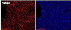
- Immunofluorescence analysis of Nanog using DyLight 550 directly conjugated anti-Nanog monoclonal antibody. Human iPSC Ad1 cells grown on vitronectin were fixed, permeabilized and stained with DyLight 550 conjugated anti-Nanog (Product # MA1-017-D550). Nuclear DNA was stained with DAPI (blue).

- Submitted by
- Invitrogen Antibodies (provider)
- Main image
- Experimental details
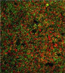
- Immunofluorescent analysis of Nanog (red) in human induced pluripotent stem cells. The cells were fixed, permeabilized and stained with a DyLight 488 conjugated SSEA3 monoclonal antibody (Product # MA1-020-D488, green) and a DyLight D550 conjugated NANOG monoclonal antibody (Product # MA1-017-D550, red) at a dilution of 1:100 in 3% BSA/PBS blocking buffer overnight at 4°C. Note: Data courtesy of Innovators Program.

- Submitted by
- Invitrogen Antibodies (provider)
- Main image
- Experimental details
- Immunofluorescence analysis of Nanog using DyLight 550 directly conjugated anti-Nanog monoclonal antibody. Human iPSC Ad1 cells grown on vitronectin were fixed, permeabilized and stained with DyLight 550 conjugated anti-Nanog (Product # MA1-017-D550). Nuclear DNA was stained with DAPI (blue).

- Submitted by
- Invitrogen Antibodies (provider)
- Main image

- Experimental details
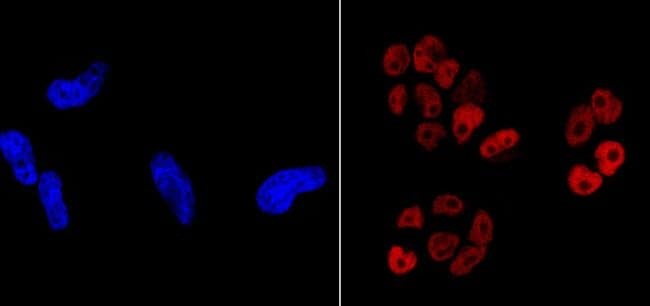
- Immunofluorescent analysis of Nanog (red) showing nuclear staining of NCCIT cells (right panel) compared to negative HeLa cell control (left panel). The cells were fixed with formalin for 15 minutes, permeabilized with 0.1% Triton X-100 in TBS, washed, and then blocked with 3% BSA-PBS for 30 minutes at room temperature. Cells were probed with a DyLight 550-conjugated Nanog monoclonal antibody (Product # MA1-017-D550) in 3% BSA-PBS at a dilution of 1:50 and incubated for 1 hour at 37C in the dark. Nuclei (left panel, blue) were stained with DAPI. Images were taken at 60X magnification.