 Explore
 Validate
 Learn
Learn Western blot
Antibody data
- Antibody Data
- Antigen structure
- References [0]
- Comments [0]
- Validations
- Western blot [1]
- Immunocytochemistry [2]
- Immunohistochemistry [2]
Submit
Validation data
Reference
Comment
Report error
- Product number
- NBP2-19458 - Provider product page

- Provider
- Novus Biologicals
- Product name
- Rabbit Polyclonal N-Cadherin Antibody
- Antibody type
- Polyclonal
- Description
- Immunogen affinity purified.
- Reactivity
- Human, Mouse
- Host
- Rabbit
- Isotype
- IgG
- Vial size
- 0.1 ml
- Storage
- Aliquot and store at -20C or -80C. Avoid freeze-thaw cycles.
No comments: Submit comment
Supportive validation
- Submitted by
- Novus Biologicals (provider)
- Main image

- Experimental details
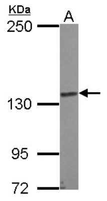
- Western Blot: N Cadherin Antibody [NBP2-19458] - Sample (30 ug of whole cell lysate) A: K562 5% SDS PAGE gel, diluted at 1:2000.
Supportive validation
- Submitted by
- Novus Biologicals (provider)
- Main image

- Experimental details
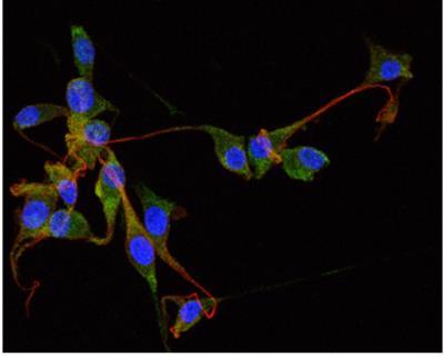
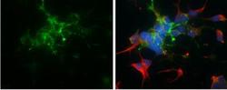
- Immunocytochemistry/Immunofluorescence: N-Cadherin Antibody [NBP2-19458] - SH-SY5Y cells were fixed in 4% paraformaldehyde at RT for 15 min. Green: N-Cadherin protein stained by N-Cadherin antibody [N1N2], N-term diluted at 1:400. Red: beta Tubulin 3/ TUJ1 protein stained by beta Tubulin 3/ TUJ1 antibody (NBP2-43559) diluted at 1:200. Blue: Hoechst 33342 staining.
- Submitted by
- Novus Biologicals (provider)
- Main image
- Experimental details
- Immunocytochemistry/Immunofluorescence: N-Cadherin Antibody [NBP2-19458] - SH-SY5Y cells were fixed in 4% paraformaldehyde at RT for 15 min.Green: N-Cadherin protein stained by N-Cadherin antibody [N1N2], N-term diluted at 1:400. Red: beta Tubulin 3/ TUJ1 protein stained by beta Tubulin 3/ TUJ1 antibody (NBP2-43559) diluted at 1:200. Blue: Hoechst 33342 staining.

Supportive validation
- Submitted by
- Novus Biologicals (provider)
- Main image

- Experimental details
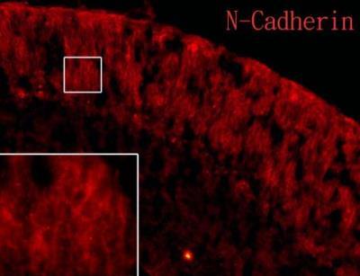
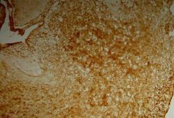
- Immunohistochemistry-Frozen: N-Cadherin Antibody [NBP2-19458] - N-Cadherin antibody [N1N2] detects N-Cadherin protein on embryonic mouse brain by immunohistochemical analysis. Sample: Frozen section of embryonic mouse brain (mE18.5). Red: N-Cadherin antibody [N1N2] diluted at 1:250.
- Submitted by
- Novus Biologicals (provider)
- Main image
- Experimental details
- Immunohistochemistry-Frozen: N-Cadherin Antibody [NBP2-19458] - N-Cadherin antibody [N1N2] detects N-Cadherin protein on embryonic mouse brain by immunohistochemical analysis. Sample: Frozen section of embryonic mouse brain (mE18.5). N-Cadherin antibody [N1N2] diluted at 1:500.
