 Explore
 Validate
 Learn
Learn Immunocytochemistry
Antibody data
- Antibody Data
- Antigen structure
- References [1]
- Comments [0]
- Validations
- Immunocytochemistry [2]
Submit
Validation data
Reference
Comment
Report error
- Product number
- MA5-17043 - Provider product page

- Provider
- Invitrogen Antibodies
- Product name
- CD3 Monoclonal Antibody (4D10A6)
- Antibody type
- Monoclonal
- Antigen
- Purifed from natural sources
- Description
- MA5-17043 targets CD3 in ICC and IF applications and shows reactivity with Human samples.
- Antibody clone number
- 4D10A6
- Concentration
- 1.0 mg/mL
Submitted references AXL Is a Key Factor for Cell Plasticity and Promotes Metastasis in Pancreatic Cancer.
Du W, Phinney NZ, Huang H, Wang Z, Westcott J, Toombs JE, Zhang Y, Beg MS, Wilkie TM, Lorens JB, Brekken RA
Molecular cancer research : MCR 2021 Aug;19(8):1412-1421
Molecular cancer research : MCR 2021 Aug;19(8):1412-1421
No comments: Submit comment
Supportive validation
- Submitted by
- Invitrogen Antibodies (provider)
- Main image
- Experimental details
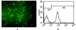
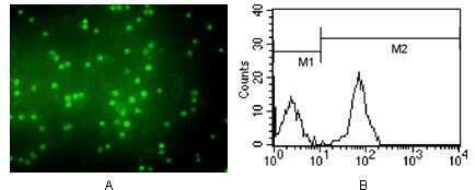
- Immunofluorescence analysis of peripheral blood T cells using CD3e/TCRE monoclonal antibody (Product # MA5-17043) (A). Flow cytometric analysis of eripheral blood T cells using CD3e/TCRE monoclonal antibody (Product # MA5-17043) (B).
- Submitted by
- Invitrogen Antibodies (provider)
- Main image
- Experimental details
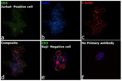
- Immunofluorescence analysis of CD3 was performed using 70% confluent log phase Jurkat cells. The cells were fixed with 4% paraformaldehyde for 10 minutes, permeabilized with 0.1% Triton™ X-100 for 15 minutes, and blocked with 2% BSA for 45 minutes at room temperature. The cells were labeled with CD3 Monoclonal Antibody (4D10A6) (Product # MA5-17043) at 1:100 dilution in 0.1% BSA, incubated at 4 degree celsius overnight and then labeled with Goat anti-Mouse IgG (H+L) Superclonal™ Recombinant Secondary Antibody, Alexa Fluor® 488 conjugate (Product # A28175), (1:2000 dilution), for 45 minutes at room temperature (Panel a: Green). Nuclei (Panel b: Blue) were stained with ProLong™ Diamond Antifade Mountant with DAPI (Product # P36962). F-actin (Panel c: Red) was stained with Rhodamine Phalloidin (Product # R415, 1:300). Panel d represents the merged image showing membrane localization. Panel e represents Raji showing no expression of CD3. Panel f represents control cells with no primary antibody to assess background. The images were captured at 60X magnification.
