Explore
 Validate
 Learn
LearnPA1-16876
antibody from Invitrogen Antibodies
Targeting: FOXP3
AIID, DIETER, IPEX, JM2, PIDX, SCURFIN, XPID
 Western blot
Antibody data
- Antibody Data
- Antigen structure
- References [0]
- Comments [0]
- Validations
- Western blot [3]
- Immunocytochemistry [1]
- Immunohistochemistry [3]
Submit
Validation data
Reference
Comment
Report error
- Product number
- PA1-16876 - Provider product page

- Provider
- Invitrogen Antibodies
- Product name
- FOXP3 Polyclonal Antibody
- Antibody type
- Polyclonal
- Antigen
- Synthetic peptide
- Description
- This antibody does not detect mouse FOXP3 in Western blot but does detect mouse tissue for IHC.
- Concentration
- 1 mg/mL
No comments: Submit comment
Supportive validation
- Submitted by
- Invitrogen Antibodies (provider)
- Main image
- Experimental details
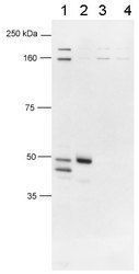
- Detection of human FOXP3 using Product # PA1-16876. Lane 1: Human CD4+CD25+ PBL, lane 2: HEK293T transfected with human Foxp3 cDNA, lane 3: 293/mouse foxp3, lane 4: 293/empty vector

- Submitted by
- Invitrogen Antibodies (provider)
- Main image

- Experimental details
- Western blot analysis of FOXP3 in human CD4+CD25+ PBL, HEK293T transfected with human Foxp3 cDNA, 293/mouse foxp3, 293/empty vector. Samples were incubated in FOXP3 polyclonal antibody (Product # PA1-16876). Lane 1: Human CD4+CD25+ PBL, lane 2: HEK293T transfected with human Foxp3 cDNA, lane 3: 293/mouse foxp3, lane 4: 293/empty vector.

- Submitted by
- Invitrogen Antibodies (provider)
- Main image
- Experimental details
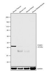
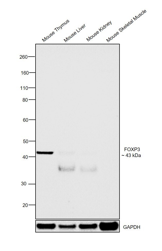
- Western blot was performed using Anti-FOXP3 Polyclonal Antibody (Product # PA1-16876) and a 43 kDa band corresponding to FOXP3 was observed in Mouse Thymus in comparison to Mouse Liver, Mouse Kidney and Mouse Skeletal Muscle which are reported to be negative. Tissue extracts (30 µg lysate) of Mouse Thymus (Lane 1), Mouse Liver (Lane 2), Mouse Kidney (Lane 3) and Mouse Skeletal Muscle (Lane 4) were electrophoresed using NuPAGE™ 4-12% Bis-Tris Protein Gel (Product # NP0322BOX). Resolved proteins were then transferred onto a nitrocellulose membrane (Product # IB23001) by iBlot® 2 Dry Blotting System (Product # IB21001). The blot was probed with the primary antibody (1:2000 dilution) and detected by chemiluminescence with Goat anti-Rabbit IgG (H+L) Superclonal™ Recombinant Secondary Antibody, HRP (Product # A27036) using the iBright FL 1000 (Product # A32752). Chemiluminescent detection was performed using SuperSignal™ West Dura Extended Duration Substrate (Product # 34076).
Supportive validation
- Submitted by
- Invitrogen Antibodies (provider)
- Main image

- Experimental details
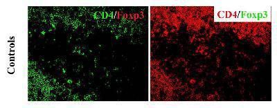
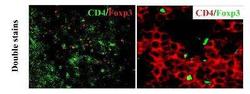
- Immunofluorescent analysis of FOXP3 incubated with CD4 and no Foxp3 using a polyclonal antibody (Product # PA1-16876).
Supportive validation
- Submitted by
- Invitrogen Antibodies (provider)
- Main image
- Experimental details
- Immunohistochemical analysis of FOXP3 in healthy thymus from a C57BL/6 mouse. Frozen samples were incubated in FOXP3 polyclonal antibody (Product # PA1-16876).

- Submitted by
- Invitrogen Antibodies (provider)
- Main image
- Experimental details
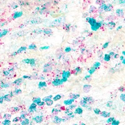
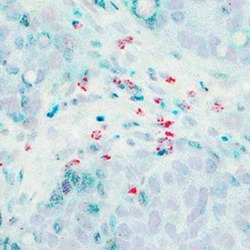
- Immunohistochemical analysis of FOXP3 in formalin-fixed paraffin-embedded tissue sections of human breast cancer. Samples were incubated in FOXP3 polyclonal antibody (Product # PA1-16876) using a dilution of 1:100. ACDs Integrated Co-Detection Workflow was performed using ACD RNAScope Probe. Tissue was stained on Leica Bond RX using RNAscope (TM) 2.5 LS Reagent Kit-RED, BOND Polymer Refine Detection (DAB) and Hematoxylin, BOND Polymer Refine Red Detection and Hematoxylin and RNAscope (TM) 2.5 LS Green Accessory Pack. Tissue was counterstained with 50% hematoxylin (blue). CD8A mRNA (red) and FOXP3 protein (green).

- Submitted by
- Invitrogen Antibodies (provider)
- Main image
- Experimental details
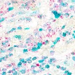
- Immunohistochemical analysis of FOXP3 in formalin-fixed paraffin-embedded tissue sections of human breast cancer. Samples were incubated in FOXP3 polyclonal antibody (Product # PA1-16876) using a dilution of 1:200. ACDs Integrated Co-Detection Workflow was performed using ACD RNAScope Probe. Tissue was stained on Leica Bond RX using RNAscope (TM) 2.5 LS Reagent Kit-RED, BOND Polymer Refine Detection (DAB) and Hematoxylin, BOND Polymer Refine Red Detection and Hematoxylin and RNAscope (TM) 2.5 LS Green Accessory Pack. Tissue was counterstained with 50% hematoxylin (blue). CD4 mRNA (red) and FOXP3 protein (green).