Explore
 Validate
 Learn
Learn Western blot
Antibody data
- Antibody Data
- Antigen structure
- References [0]
- Comments [0]
- Validations
- Western blot [2]
- Immunocytochemistry [2]
- Immunohistochemistry [7]
Submit
Validation data
Reference
Comment
Report error
- Product number
- PA5-54779 - Provider product page

- Provider
- Invitrogen Antibodies
- Product name
- PCM1 Polyclonal Antibody
- Antibody type
- Polyclonal
- Antigen
- Recombinant full-length protein
- Description
- Immunogen sequence: TIYSEVATLI SQNESRPHFL IELFHELQLL NTDYLRQRAL YALQDIVSRH ISESHEKGEN VKSVNSGTWI ASNSELTPSE SLATTDDETF EKNFE
- Concentration
- 0.1 mg/mL
No comments: Submit comment
Supportive validation
- Submitted by
- Invitrogen Antibodies (provider)
- Main image
- Experimental details
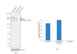
- Knockdown of PCM1 was achieved by transfecting A549 with PCM1 specific siRNAs (Silencer® select Product # S10128, S10127). Western blot analysis (Fig. a) was performed using Whole cell extracts from the PCM1 knockdown cells (lane 3), non-targeting scrambled siRNA transfected cells (lane 2) and untransfected cells (lane 1). The blot was probed with PCM1 Polyclonal Antibody (Product # PA5-54779, 1:500 dilution) and Goat anti-Rabbit IgG (H+L) Superclonal™ Recombinant Secondary Antibody, HRP (Product # A27036, 1:4000 dilution). Densitometric analysis of this western blot is shown in histogram (Fig. b). Decrease in signal upon siRNA mediated knock down confirms that antibody is specific to PCM1.

- Submitted by
- Invitrogen Antibodies (provider)
- Main image

- Experimental details
- Western blot was performed using Anti-PCM1 Polyclonal Antibody (Product # PA5-54779) and a 250kDa and 225kDa band corresponding to PCM1 were observed across cell lines and tissue tested. Whole cell extracts (40 µg lysate) of HEK-293 (Lane 1), U-2 OS (Lane 2), NIH/3T3 (Lane 3), A549 (Lane 4) and K-562 (Lane 5) as seen in Fig (a). Tissue extracts of Mouse Testis (Lane 1) and Rat Testis (Lane 2) as seen in Fig (b) were electrophoresed using NuPAGE™ 3-8% Tris-Acetate Protein Gel (Product # EA0378BOX). Resolved proteins were then transferred onto a Nitrocellulose membrane (Product # IB23002) by iBlot® 2 Dry Blotting System (Product # IB21001) and then equilibrated with 20% ethanol. The blot was probed with the primary antibody (1:500 dilution) and detected by chemiluminescence with Goat anti-Rabbit IgG (H+L) Superclonal™ Recombinant Secondary Antibody, HRP (Product # A27036, 1:4000 dilution) using the iBright FL 1000 (Product # A32752). Chemiluminescent detection was performed using Novex® ECL Chemiluminescent Substrate Reagent Kit (Product # WP20005).

Supportive validation
- Submitted by
- Invitrogen Antibodies (provider)
- Main image

- Experimental details
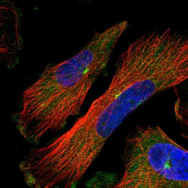
- Immunofluorescent staining of PCM1 in human cell line U-251 MG shows positivity in nuclear membrane & centrosome. Samples were probed using a PCM1 Polyclonal Antibody (Product # PA5-54779).
- Submitted by
- Invitrogen Antibodies (provider)
- Main image

- Experimental details
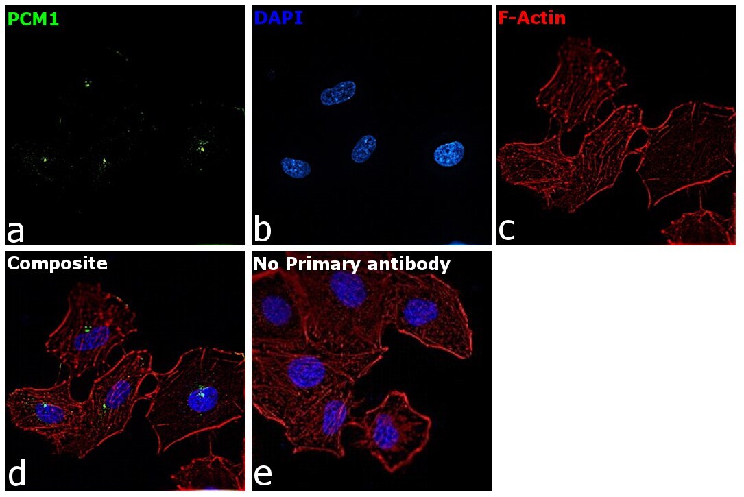
- Immunofluorescence analysis of PCM1 was performed using 70% confluent log phase A549 cells. The cells were fixed with 4% paraformaldehyde for 10 minutes, permeabilized with 0.1% Triton™ X-100 for 15 minutes, and blocked with 2% BSA for 45 minutes at room temperature. The cells were labeled with PCM1 Polyclonal Antibody (Product # PA5-54779) at 1:100 dilution in 0.1% BSA, incubated at 4 degree celsius overnight and then labeled with Donkey anti-Rabbit IgG (H+L) Highly Cross-Adsorbed Secondary Antibody, Alexa Fluor Plus 488 (Product # A32790), (1:2000 dilution), for 45 minutes at room temperature (Panel a: Green). Nuclei (Panel b:Blue) were stained with ProLong™ Diamond Antifade Mountant with DAPI (Product # P36962). F-actin (Panel c: Red) was stained with Rhodamine Phalloidin (Product # R415, 1:300 dilution). Panel d represents the merged image showing centrosomal localization. Panel e represents control cells with no primary antibody to assess background. The images were captured at 60X magnification.
Supportive validation
- Submitted by
- Invitrogen Antibodies (provider)
- Main image
- Experimental details
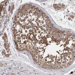
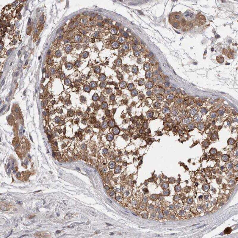
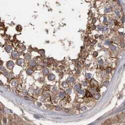
- Immunohistochemical staining of PCM1 in human testis using a PCM1 Polyclonal Antibody (Product # PA5-54779) shows moderate to strong cytoplasmic positivity in cells in seminiferous ducts.
- Submitted by
- Invitrogen Antibodies (provider)
- Main image
- Experimental details
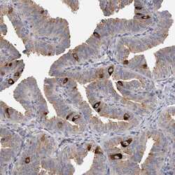
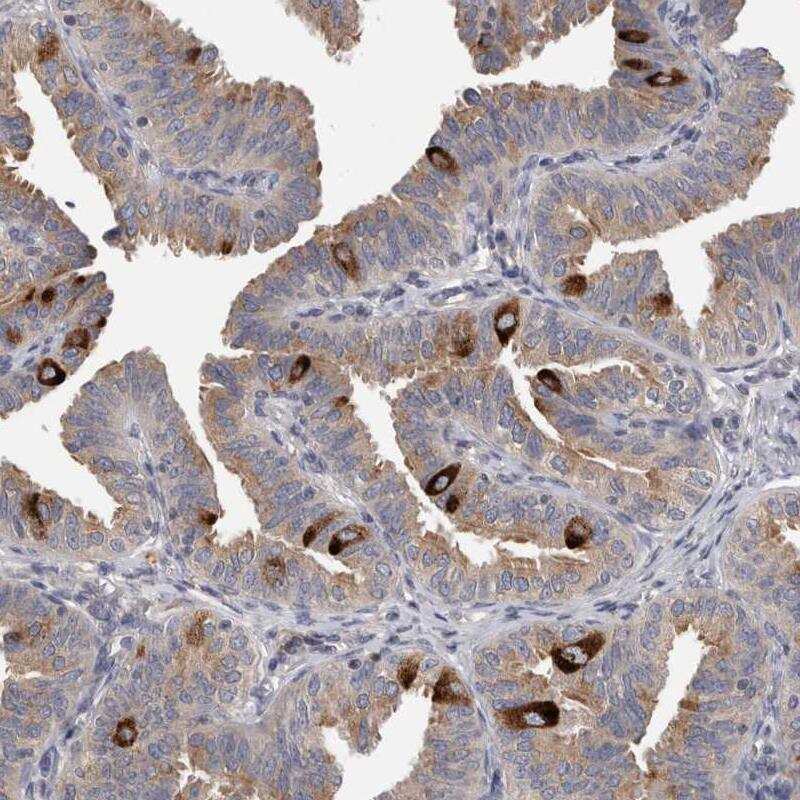
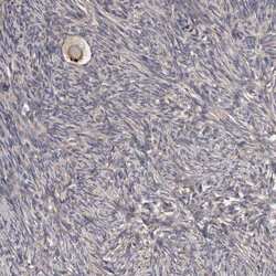
- Immunohistochemical staining of PCM1 in human fallopian tube using a PCM1 Polyclonal Antibody (Product # PA5-54779) shows strong cytoplasmic positivity in a subset of glandular cells.
- Submitted by
- Invitrogen Antibodies (provider)
- Main image
- Experimental details
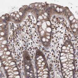
- Immunohistochemical staining of PCM1 in human rectum using a PCM1 Polyclonal Antibody (Product # PA5-54779) shows moderate cytoplasmic positivity in glandular cells.

- Submitted by
- Invitrogen Antibodies (provider)
- Main image
- Experimental details
- Immunohistochemical staining of PCM1 in human ovary using a PCM1 Polyclonal Antibody (Product # PA5-54779) shows strong positivity in oocyte.

- Submitted by
- Invitrogen Antibodies (provider)
- Main image
- Experimental details
- Immunohistochemical staining of PCM1 in human testis using PCM1 Polyclonal Antibody (Product # PA5-54779).

- Submitted by
- Invitrogen Antibodies (provider)
- Main image
- Experimental details
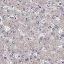
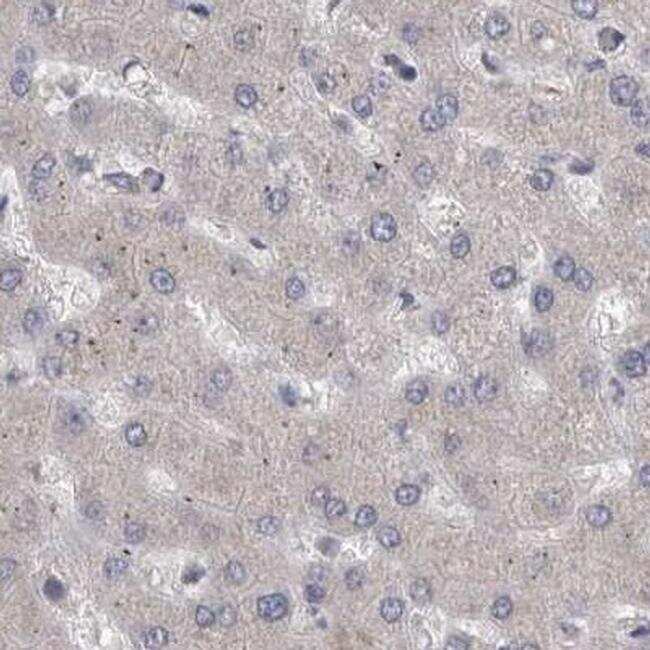
- Immunohistochemical staining of PCM1 in human liver using PCM1 Polyclonal Antibody (Product # PA5-54779).
- Submitted by
- Invitrogen Antibodies (provider)
- Main image
- Experimental details
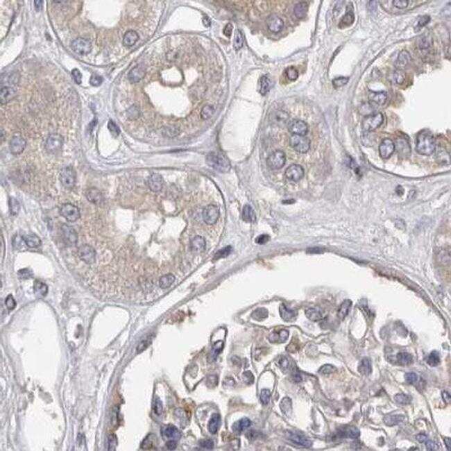
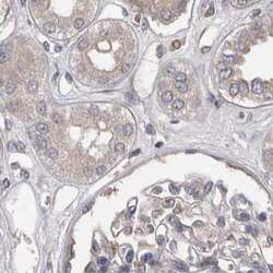
- Immunohistochemical staining of PCM1 in human kidney using PCM1 Polyclonal Antibody (Product # PA5-54779).