Explore
 Validate
 Learn
LearnPA1-32079
antibody from Invitrogen Antibodies
Targeting: ATP6AP2
APT6M8-9, ATP6IP2, ATP6M8-9, M8-9, PRR, RENR
 Western blot
 Immunohistochemistry
Antibody data
- Antibody Data
- Antigen structure
- References [0]
- Comments [0]
- Validations
- Western blot [4]
- Flow cytometry [1]
Submit
Validation data
Reference
Comment
Report error
- Product number
- PA1-32079 - Provider product page

- Provider
- Invitrogen Antibodies
- Product name
- ATP6IP2 Polyclonal Antibody
- Antibody type
- Polyclonal
- Antigen
- Synthetic peptide
- Description
- Recommended positive controls: The peptide used to generate this antibody is available for purchase (GTX25959-PEP)..
- Reactivity
- Human, Mouse, Rat
- Host
- Goat
- Isotype
- IgG
- Vial size
- 50 µg
- Concentration
- 0.5 mg/mL
- Storage
- Store at 4°C short term. For long term storage, store at -20°C, avoiding freeze/thaw cycles.
No comments: Submit comment
Supportive validation
- Submitted by
- Invitrogen Antibodies (provider)
- Main image
- Experimental details
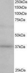
- Western blot analysis of ATP6IP2 in human Kidney lysate (RIPA buffer 35 µg total protein per lane) using an ATP6IP2 polyclonal antibody (Product # PA1-32079) at a dilution of 0.5 µg/mL following incubation for 1 hour and detected using chemiluminescence.
- Submitted by
- Invitrogen Antibodies (provider)
- Main image

- Experimental details
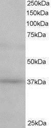
- Western Blot analysis of ATP6IP2 was performed by loading 35 µg (in RIPA buffer) of mouse kidney lysates. Proteins were transferred to a membrane and probed with a ATP6IP2 Polyclonal Antibody (Product # PA1-32079) at a dilution of 2 µg/mL.
- Submitted by
- Invitrogen Antibodies (provider)
- Main image
- Experimental details
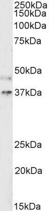
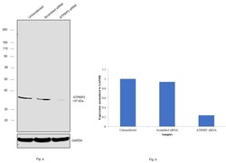
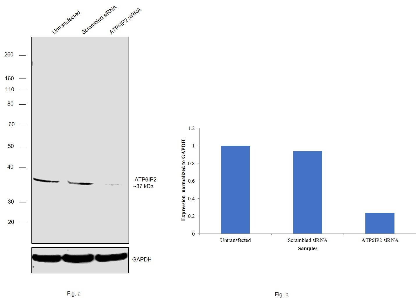
- Knockdown of ATP6IP2 was achieved by transfecting MCF-7 cells with ATP6IP2 specific siRNAs (Silencer® select Product # s19791, s19790). Western blot analysis (Fig. a) was performed using whole cell extracts from the MCF-7 knockdown cells (lane 3), non-specific scrambled siRNA transfected cells (lane 2) and untransfected cells (lane 1). The blot was probed with ATP6IP2 Polyclonal Antibody (Product # PA1-32079, 0.125 µg/mL) and Rabbit anti-Goat IgG (H+L), Superclonal™ Recombinant Secondary Antibody, HRP (Product # A27014, 1:4000 dilution). Densitometric analysis of this western blot is shown in histogram (Fig. b, Fig.c). Decrease in signal upon siRNA mediated knock down confirms that antibody is specific to ATP6IP2.
- Submitted by
- Invitrogen Antibodies (provider)
- Main image
- Experimental details
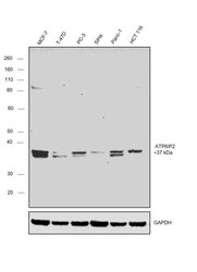
- Western blot was performed using Anti-ATP6IP2 Polyclonal Antibody (Product # PA1-32079) and ~37 kDa band corresponding to ATP6IP2 was observed across the panel tested. Whole cell extracts (30 µg lysate) of MCF-7 (Lane 1), T-47D (Lane 2), PC-3 (Lane 3), SiHa (Lane 4), Panc-1 (Lane 5) and HCT 116 (Lane 6) were electrophoresed using NuPAGE™ 10% Bis-Tris Protein Gel (Product # NP0302BOX). Resolved proteins were then transferred onto a nitrocellulose membrane (Product # IB23001) by iBlot® 2 Dry Blotting System (Product # IB21001). The blot was probed with the primary antibody (0.5 µg/mL) and detected by chemiluminescence with Rabbit anti-Goat IgG (H+L), Superclonal™ Recombinant Secondary Antibody, HRP (Product # A27014, 1:4000 dilution) using the iBright FL 1000 (Product # A32752). Chemiluminescent detection was performed using Novex® ECL Chemiluminescent Substrate Reagent Kit (Product # WP20005).

Supportive validation
- Submitted by
- Invitrogen Antibodies (provider)
- Main image
- Experimental details
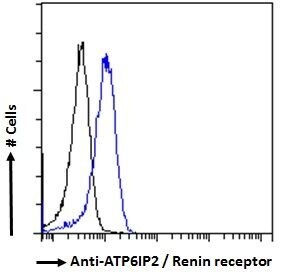
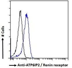
- Flow Cytometry analysis of ATP6V1A was performed in HeLa cells (fixed with PFA, permeabilized with 0.5% Triton) using a ATP6V1A Monoclonal Antibody (GT1561) (Product # MA5-31554) (Blue) at a dilution of 10 µg/mL. Black : Isotype control.