Explore
 Validate
 Learn
Learn Western blot
 ELISA
ELISA Immunohistochemistry
Antibody data
- Antibody Data
- Antigen structure
- References [0]
- Comments [0]
- Validations
- Immunohistochemistry [4]
Submit
Validation data
Reference
Comment
Report error
- Product number
- LS-C745260 - Provider product page

- Provider
- LSBio
- Product name
- IDO1 / IDO Antibody (clone 2E2.6) LS-C745260
- Antibody type
- Monoclonal
- Description
- Protein G affinity chromatography
- Reactivity
- Mouse
- Host
- Mouse
- Isotype
- IgG
- Antibody clone number
- 2E2.6
- Storage
- Store vial at -20°C or below prior to opening. Dilute 1:10 to minimize loss. Store the vial at -20°C or below after dilution. Avoid freeze-thaw cycles.
No comments: Submit comment
Supportive validation
- Submitted by
- LSBio (provider)
- Enhanced method
- Genetic validation
- Main image

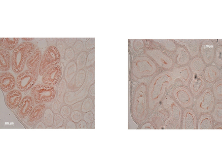
- Experimental details
- Immunohistochemistry of Mouse anti-IDO1 antibody. Tissue: epididymis from wild-type (left) or IDO1 null mice (right). Fixation: paraffin-embedded. Primary antibody: IDO1 (2E2) monoclonal antibody. Secondary antibody: Peroxidase mouse secondary antibody at 1:10,000 for 45 min at RT. Localization: IDO-1 is located in the cytosol. Staining: IDO1 as precipitated brown signal.

- Submitted by
- LSBio (provider)
- Enhanced method
- Genetic validation
- Main image

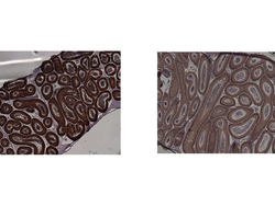
- Experimental details
- Immunohistochemistry of Mouse Anti-IDO1 Antibody. Tissue: epididymis from wild-type (left) or IDO1 null mice (right). Fixation: frozen sections. Antigen retrieval: not required. Primary antibody: IDO1 (2E2) monoclonal antibody. Secondary antibody: Peroxidase mouse secondary antibody at 1:10,000 for 45 min at RT. Localization: IDO-1 is located in the cytosol. Staining: IDO-1 as precipitated brown signal.

- Submitted by
- LSBio (provider)
- Main image
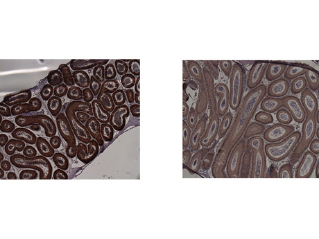
- Experimental details
- Immunohistochemistry of Mouse anti-IDO1 antibody. Tissue: epididymis from wild-type (left) or IDO1 null mice (right). Fixation: paraffin-embedded. Primary antibody: IDO1 (2E2) monoclonal antibody. Secondary antibody: Peroxidase mouse secondary antibody at 1:10,000 for 45 min at RT. Localization: IDO-1 is located in the cytosol. Staining: IDO1 as precipitated brown signal.
- Submitted by
- LSBio (provider)
- Main image

- Experimental details
- Immunohistochemistry of Mouse Anti-IDO1 Antibody. Tissue: epididymis from wild-type (left) or IDO1 null mice (right). Fixation: frozen sections. Antigen retrieval: not required. Primary antibody: IDO1 (2E2) monoclonal antibody. Secondary antibody: Peroxidase mouse secondary antibody at 1:10,000 for 45 min at RT. Localization: IDO-1 is located in the cytosol. Staining: IDO-1 as precipitated brown signal.