Explore
 Validate
 Learn
Learn Western blot
Antibody data
- Antibody Data
- Antigen structure
- References [2]
- Comments [0]
- Validations
- Western blot [2]
- Immunocytochemistry [1]
- Flow cytometry [1]
Submit
Validation data
Reference
Comment
Report error
- Product number
- 44-536G - Provider product page

- Provider
- Invitrogen Antibodies
- Product name
- Phospho-HSP25 (Ser86) Polyclonal Antibody
- Antibody type
- Polyclonal
- Antigen
- Synthetic peptide
- Reactivity
- Human, Mouse
- Host
- Rabbit
- Isotype
- IgG
- Vial size
- 100 µL
- Storage
- -20°C
Submitted references PFKFB3 activation in cancer cells by the p38/MK2 pathway in response to stress stimuli.
p38 MAPK/HSP25 signaling mediates cadmium-induced contraction of mesangial cells and renal glomeruli.
Novellasdemunt L, Bultot L, Manzano A, Ventura F, Rosa JL, Vertommen D, Rider MH, Navarro-Sabate À, Bartrons R
The Biochemical journal 2013 Jun 15;452(3):531-43
The Biochemical journal 2013 Jun 15;452(3):531-43
p38 MAPK/HSP25 signaling mediates cadmium-induced contraction of mesangial cells and renal glomeruli.
Hirano S, Sun X, DeGuzman CA, Ransom RF, McLeish KR, Smoyer WE, Shelden EA, Welsh MJ, Benndorf R
American journal of physiology. Renal physiology 2005 Jun;288(6):F1133-43
American journal of physiology. Renal physiology 2005 Jun;288(6):F1133-43
No comments: Submit comment
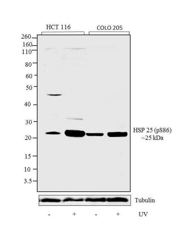
Supportive validation
- Submitted by
- Invitrogen Antibodies (provider)
- Main image

- Experimental details
- Western blot analysis was performed on whole cell extracts (30 µg lysate) of HCT 116 (Lane 1), HCT 116 treated with UV for 40 minutes (lane 2), COLO 205 (lane 3), and COLO 205 treated with UV for 40 minutes (Lane 4). The blots were probed with Anti-Phospho-HSP 25 pSer86 Rabbit Polyclonal Antibody (Product # 44-536G, 1 in 1000 dilution) and detected by chemiluminescence Goat Anti-Rabbit IgG Secondary Antibody, HRP conjugate (Product # G-21234, 1:5000 dilution). A 25 kDa band corresponding to Phospho-HSP25 pSer86 was observed across cell lines tested. Known quantity of protein samples were electrophoresed using Novex® NuPAGE® 10 % Bis-Tris gel (Product # NP0301BOX), XCell SureLock™ Electrophoresis System (Product # EI0002) and Novex® Sharp Pre-Stained Protein Standard (Product # LC5800). Resolved proteins were then transferred onto a nitrocellulose membrane by iBlot® 2 Dry Blotting System (Product # IB21001).The membrane was probed with the relevant primary and secondary Antibody following blocking with 5 % skimmed milk. Chemiluminescent detection was performed using Pierce™ ECL Western Blotting Substrate (Product # 32106).
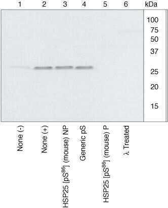
- Submitted by
- Invitrogen Antibodies (provider)
- Main image
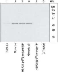
- Experimental details
- HSP25 (pS86) phosphospecific antibody. Peptide competition shows that only the peptide corresponding to HSP25 (pS86) blocks antibody signal, thereby demonstrating the specificity of the antibody.
Supportive validation
- Submitted by
- Invitrogen Antibodies (provider)
- Main image
- Experimental details
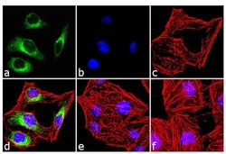
- Immunofluorescence analysis of Phospho-Hsp25 pSer86 was done on 70% confluent log phase HeLa cells treated with UV-302nm for 60 minutes. The cells were fixed with 4% paraformaldehyde for 10 minutes, permeabilized with 0.1% Triton™ X-100 for 10 minutes, and blocked with 1% BSA for 1 hour at room temperature. The cells were labeled with Phospho-Hsp25 pSer86 Rabbit Polyclonal Antibody (Product # 44-536G) at 1:250 dilution in 0.1% BSA and incubated for 3 hours at room temperature and then labeled with Goat anti-Rabbit IgG (H+L) Superclonal™ Secondary Antibody, Alexa Fluor® 488 conjugate (Product # A27034) at a dilution of 1:2000 for 45 minutes at room temperature (Panel a: green). Nuclei (Panel b: blue) were stained with SlowFade® Gold Antifade Mountant with DAPI (Product # S36938). F-actin (Panel c: red) was stained with Rhodamine Phalloidin (Product # R415, 1:300). Panel d is a merged image showing cytoplasmic localization. Panel e is untreated cell with no signal. Panel f is a no primary antibody control. The images were captured at 60X magnification.
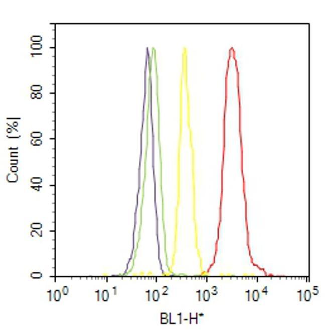
Supportive validation
- Submitted by
- Invitrogen Antibodies (provider)
- Main image
- Experimental details
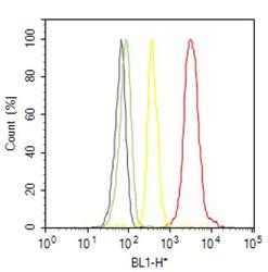
- Flow cytometry analysis of Phospho-Hsp25 [pSer86] was done on HeLa cells treated with UV (30 minutes). Cells were fixed with 70% ethanol for 10 minutes, permeabilized with 0.25% Triton™ X-100 for 20 minutes, and blocked with 5% BSA for 30 minutes at room temperature. Cells were labeled with Phospho-Hsp25 [pSer86] Rabbit Polyclonal Antibody (44536G, red histogram) or with rabbit isotype control (yellow histogram) at 3-5 ug/million cells in 2.5% BSA. After incubation at room temperature for 2 hours, the cells were labeled with Alexa Fluor® 488 Goat Anti-Rabbit Secondary Antibody (A11008) at a dilution of 1:400 for 30 minutes at room temperature. The representative 10,000 cells were acquired and analyzed for each sample using an Attune® Acoustic Focusing Cytometer. The purple histogram represents unstained control cells and the green histogram represents no-primary-antibody control..