Explore
 Validate
 Learn
Learn Western blot
 Immunocytochemistry
Antibody data
- Antibody Data
- Antigen structure
- References [0]
- Comments [0]
- Validations
- Western blot [2]
- Flow cytometry [1]
Submit
Validation data
Reference
Comment
Report error
- Product number
- MAB9355 - Provider product page

- Provider
- Novus Biologicals
- Product name
- Rabbit Monoclonal HSP27 Antibody
- Antibody type
- Monoclonal
- Description
- Protein A or G purified.
- Reactivity
- Human
- Host
- Rabbit
- Isotype
- IgG
- Vial size
- 100 ug
- Concentration
- 1.0 mg/ml
- Storage
- Store at -20C. Avoid freeze-thaw cycles.
No comments: Submit comment
Supportive validation
- Submitted by
- Novus Biologicals (provider)
- Main image
- Experimental details
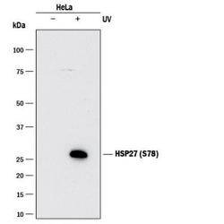
- Western Blot: HSP27 [p Ser78] Antibody (2079A) [MAB9355] - Detection of Human HSP27 [p Ser78] by Western Blot. Western blot shows lysates of HeLa human cervical epithelial carcinoma cell line untreated (-) or UV treated (+). PVDF membrane was probed with Rabbit Anti-Human HSP27 [p Ser78] Monoclonal Antibody (Catalog # MAB9355) followed by HRP-conjugated Anti-Rabbit IgG Secondary Antibody. A specific band was detected for HSP27 [p Ser78 at approximately 27 kDa (as indicated). This experiment was conducted under reducing conditions

- Submitted by
- Novus Biologicals (provider)
- Main image
- Experimental details
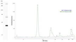
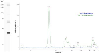
- Simple Western: HSP27 [p Ser78] Antibody (2079A) [MAB9355] - Lane vies shows a specific band for HSP27 using 0.2 mg/mL MCF-7 -/+ UV cell lysates and antibody at 5 ug/mL. Electropherogram image of corresponding Simple Western lanes view at WES molecular weight of 32 kDa. Image reported by internal validation.
Supportive validation
- Submitted by
- Novus Biologicals (provider)
- Main image

- Experimental details
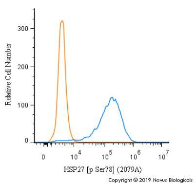
- Flow Cytometry: HSP27 [p Ser78] Antibody (2079A) [MAB9355] - An intracellular stain was performed on MCF7 cells with HSP27 [p Ser78] Antibody MAB9355 (blue) and a matched isotype control (orange). Cells were fixed with 4% PFA and then permeabilized with 0.1% saponin. Cells were incubated in an antibody dilution of 1.0 ug/mL for 30 minutes at room temperature, followed by Rabbit IgG (H+L) Cross-Adsorbed Secondary Antibody, Dylight 550 (SA5-10033, Thermo Fisher).