 Explore
 Validate
 Learn
Learn Western blot
Antibody data
- Antibody Data
- Antigen structure
- References [3]
- Comments [0]
- Validations
- Western blot [3]
- Immunocytochemistry [4]
- Immunohistochemistry [2]
- Other assay [2]
Submit
Validation data
Reference
Comment
Report error
- Product number
- PA1-017 - Provider product page

- Provider
- Invitrogen Antibodies
- Product name
- HSP27 Polyclonal Antibody
- Antibody type
- Polyclonal
- Antigen
- Synthetic peptide
- Description
- PA1-017 detects heat shock protein 27(hsp27) from rat, mouse, monkey, and human tissue tissues.
- Concentration
- 1 mg/mL
Submitted references Heat Shock Protein A6 Is Especially Involved in Enterovirus 71 Infection.
The Glyoxalase System Is a Novel Cargo of Amniotic Fluid Stem-Cell-Derived Extracellular Vesicles.
Small heat shock protein 27 (Hsp27) expression is highly induced in rat myometrium during late pregnancy and labour.
Jia J, Liu G, Zhong J, Yan R, Song X, Zheng K, Ren Z, He Z, Zhu Q
Frontiers in microbiology 2022;13:865644
Frontiers in microbiology 2022;13:865644
The Glyoxalase System Is a Novel Cargo of Amniotic Fluid Stem-Cell-Derived Extracellular Vesicles.
Romani R, Talesa VN, Antognelli C
Antioxidants (Basel, Switzerland) 2022 Aug 5;11(8)
Antioxidants (Basel, Switzerland) 2022 Aug 5;11(8)
Small heat shock protein 27 (Hsp27) expression is highly induced in rat myometrium during late pregnancy and labour.
White BG, Williams SJ, Highmore K, Macphee DJ
Reproduction (Cambridge, England) 2005 Jan;129(1):115-26
Reproduction (Cambridge, England) 2005 Jan;129(1):115-26
No comments: Submit comment
Supportive validation
- Submitted by
- Invitrogen Antibodies (provider)
- Main image

- Experimental details
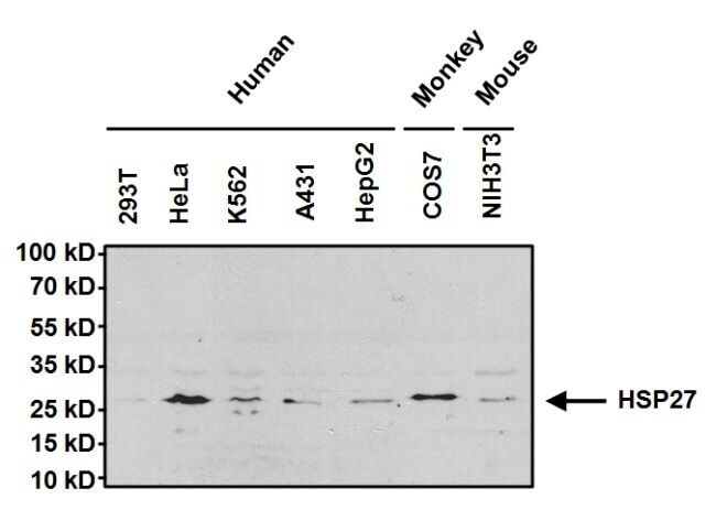
- Western blot analysis of Heat Shock Protein 27 (HSP27) was performed by loading 50 µg of the indicated whole cell lysates per well onto a 4-20% Tris-HCl polyacrylamide gel. Proteins were transferred to a PVDF membrane and blocked with 5% BSA/TBST for at least 1 hour. The membrane was probed with a HSP27 polyclonal antibody (Product # PA1-017) at a dilution of 1:1000 overnight at 4°C on a rocking platform, washed in TBS-0.1%Tween 20, and probed with a goat anti-rabbit IgG-HRP secondary antibody (Product # 31460) at a dilution of 1:20,000 for at least one hour. Chemiluminescent detection was performed using SuperSignal West Pico (Product # 34080).
- Submitted by
- Invitrogen Antibodies (provider)
- Main image
- Experimental details
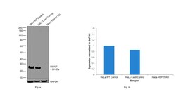
- Knockout of HSP27 was achieved by CRISPR-Cas9 genome editing using LentiArray™ Lentiviral sgRNA (Product # A32042, Assay ID CRISPR990059_LV) and LentiArray Cas9 Lentivirus (Product # A32064). Western blot analysis of HSP27 was performed by loading 30 µg of HeLa Wild type (Lane 1), HeLa Cas9 (Lane 2) and HeLa HSP27 KO (Lane 3) whole cell extracts. The samples were electrophoresed using NuPAGE™ Novex™ 4-12% Bis-Tris Protein Gel (Product # NP0321BOX). Resolved proteins were then transferred onto a nitrocellulose membrane (Product # IB23001) by iBlot® 2 Dry Blotting System (Product # IB21001). The blot was probed with Anti-HSP27 Polyclonal Antibody (Product # PA1-017, 1:2,000 dilution) and Goat anti-Rabbit IgG (H+L) Superclonal™ Recombinant Secondary Antibody, HRP (Product # A27036, 1:10,000 dilution) using the iBright FL 1000 (Product # A32752). Chemiluminescent detection was performed using Novex® ECL Chemiluminescent Substrate Reagent Kit (Product # WP20005). Loss of signal upon CRISPR mediated knockout (KO) using the LentiArray™ CRISPR product line confirms that antibody is specific to HSP27.

- Submitted by
- Invitrogen Antibodies (provider)
- Main image
- Experimental details
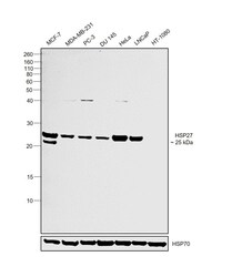
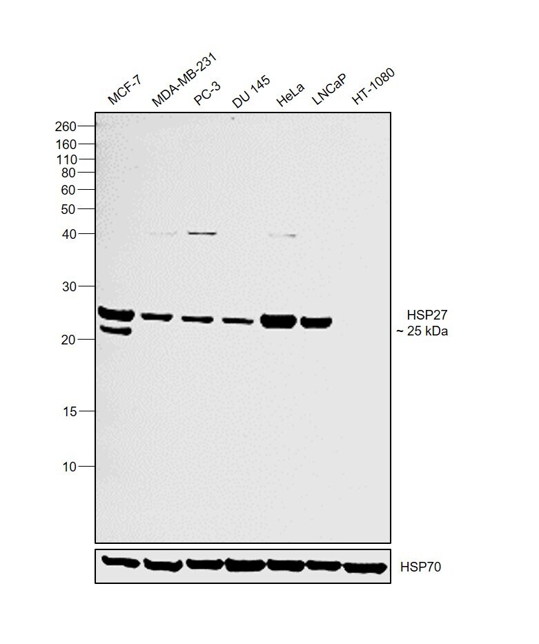
- Western blot was performed using Anti-HSP27 Polyclonal Antibody(Product # PA1-017) and a 25kDa band corresponding to HSP27 was observed across cell lines tested except HT-1080 which is reported to be negative. Whole cell extracts (30 µg lysate) of MCF7 (Lane 1), MDA-MB-231 (Lane 2), PC-3 (Lane 3), DU 145 (Lane 4), HeLa (Lane 5), LNCaP (Lane 6) and HT-1080 (Lane 7) were electrophoresed using NuPAGE™ 12% Bis-Tris Protein Gel (Product # NP0342BOX). Resolved proteins were then transferred onto a Nitrocellulose membrane (Product # IB23001) by iBlot® 2 Dry Blotting System (Product # IB21001). The blot was probed with the primary antibody (1:2000 dilution) and detected by chemiluminescence with Goat anti-Rabbit IgG (H+L) Superclonal™ Recombinant Secondary Antibody, HRP (Product # A27036,1:4000 dilution) using the iBright FL 1000 (Product # A32752). Chemiluminescent detection was performed using Novex® ECL Chemiluminescent Substrate Reagent Kit (Product # WP20005).
Supportive validation
- Submitted by
- Invitrogen Antibodies (provider)
- Main image
- Experimental details
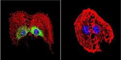
- Immunofluorescent analysis of Heat Shock Protein 27 in C6 Cells. Cells were grown on chamber slides and fixed with formaldehyde prior to staining. Cells were probed without (control) or with a Heat Shock Protein 27 polyclonal antibody (Product # PA1-017) at a dilution of 1:100 overnight at 4 C, washed with PBS and incubated with a DyLight-488 conjugated secondary antibody (Product # 35552). Heat Shock Protein 27 staining (green), F-Actin staining with Phalloidin (red) and nuclei with DAPI (blue) is shown. Images were taken at 60X magnification.
- Submitted by
- Invitrogen Antibodies (provider)
- Main image

- Experimental details
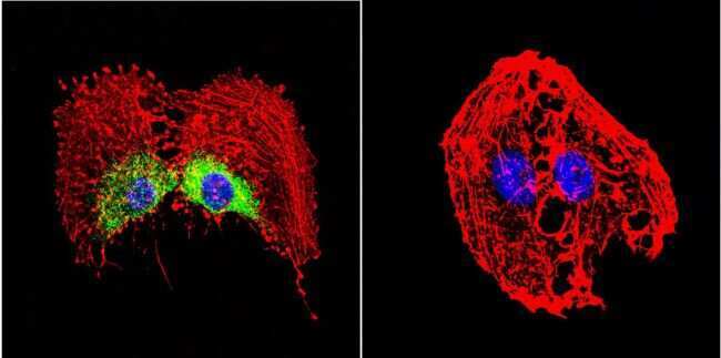
- Immunofluorescent analysis of Heat Shock Protein 27 in HeLa Cells. Cells were grown on chamber slides and fixed with formaldehyde prior to staining. Cells were probed without (control) or with a Heat Shock Protein 27 polyclonal antibody (Product # PA1-017) at a dilution of 1:200 overnight at 4 C, washed with PBS and incubated with a DyLight-488 conjugated secondary antibody (Product # 35552). Heat Shock Protein 27 staining (green), F-Actin staining with Phalloidin (red) and nuclei with DAPI (blue) is shown. Images were taken at 60X magnification.

- Submitted by
- Invitrogen Antibodies (provider)
- Main image
- Experimental details
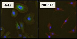
- Immunofluorescent analysis of Heat Shock Protein 27 (HSP27) (green) in HeLa cells and negative control NIH3T3 cells. Formalin fixed cells were permeabilized with 0.1% Triton X-100 in TBS for 10 minutes at room temperature and blocked with 1% Blocker BSA (Product # 37525) for 15 minutes at room temperature. Cells were probed with HSP27 polyclonal antibody (Product # PA1-017), at a dilution of 1:50 for at least 1 hour at room temperature, washed with PBS, and incubated with DyLight 488 goat-anti-rabbit IgG secondary antibody (Product # 35552) at a dilution of 1:400 for 30 minutes at room temperature. F-Actin (red) was stained with DyLight 554 Phalloidin (Product # 21834) and nuclei (blue) were stained with Hoechst 33342 dye (Product # 62249). Images were taken on a Thermo Scientific ArrayScan at 20X magnification.

- Submitted by
- Invitrogen Antibodies (provider)
- Main image

- Experimental details
- Immunofluorescent analysis of Heat Shock Protein 27 in MCF-7 Cells. Cells were grown on chamber slides and fixed with formaldehyde prior to staining. Cells were probed without (control) or with a Heat Shock Protein 27 polyclonal antibody (Product # PA1-017) at a dilution of 1:200 overnight at 4 C, washed with PBS and incubated with a DyLight-488 conjugated secondary antibody (Product # 35552). Heat Shock Protein 27 staining (green), F-Actin staining with Phalloidin (red) and nuclei with DAPI (blue) is shown. Images were taken at 60X magnification.

Supportive validation
- Submitted by
- Invitrogen Antibodies (provider)
- Main image
- Experimental details
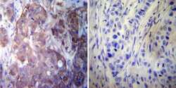
- Immunohistochemistry was performed on cancer biopsies of deparaffinized human Breast carcinoma tissue. To expose target proteins, heat induced antigen retrieval was performed using 10mM sodium citrate (pH6.0) buffer, microwaved for 8-15 minutes. Following antigen retrieval tissues were blocked in 3% BSA-PBS for 30 minutes at room temperature. Tissues were then probed at a dilution of 1:100 with a rabbit polyclonal antibody recognizing Heat Shock Protein 27 (Product # PA1-017) or without primary antibody (negative control) overnight at 4°C in a humidified chamber. Tissues were washed extensively with PBST and endogenous peroxidase activity was quenched with a peroxidase suppressor. Detection was performed using a biotin-conjugated secondary antibody and SA-HRP, followed by colorimetric detection using DAB. Tissues were counterstained with hematoxylin and prepped for mounting.

- Submitted by
- Invitrogen Antibodies (provider)
- Main image
- Experimental details
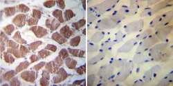
- Immunohistochemistry was performed on normal deparaffinized human Skeletal muscle tissue. To expose target proteins, heat induced antigen retrieval was performed using 10mM sodium citrate (pH6.0) buffer, microwaved for 8-15 minutes. Following antigen retrieval tissues were blocked in 3% BSA-PBS for 30 minutes at room temperature. Tissues were then probed at a dilution of 1:20 with a rabbit polyclonal antibody recognizing Heat Shock Protein 27 (Product # PA1-017) or without primary antibody (negative control) overnight at 4°C in a humidified chamber. Tissues were washed extensively with PBST and endogenous peroxidase activity was quenched with a peroxidase suppressor. Detection was performed using a biotin-conjugated secondary antibody and SA-HRP, followed by colorimetric detection using DAB. Tissues were counterstained with hematoxylin and prepped for mounting.

Supportive validation
- Submitted by
- Invitrogen Antibodies (provider)
- Main image

- Experimental details
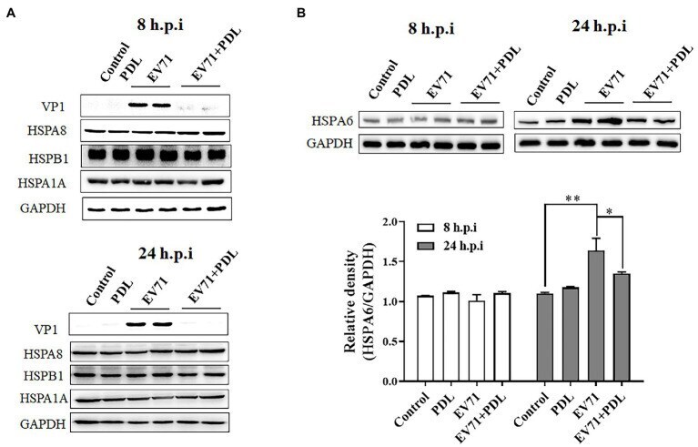
- Figure 2 EV71 infection specially upregulates HSPA6 protein expression. (A) Western blotting analysis of HSPA1A, HSPA8, HSPB1, and viral VP1 expression in RD cells infected with EV71 for 8 or 24 h. (B) Western blotting analysis of HSPA6 expression in RD cells infected with EV71 for 8 or 24 h. The relative expression of HSPA6 was quantitatively analyzed based on the bands' intensities from the Western blotting analysis. PDL (10 muM) here was used as anti-EV71 control ( * p < 0.05, ** p < 0.01).
- Submitted by
- Invitrogen Antibodies (provider)
- Main image
- Experimental details
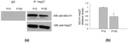
- Heat shock protein (Hsp)27 is a MG-H1-modified protein in HASC-P10 and HASC-P100 EVs. ( a ) Lysates from HASC-P10 and HASC-P100 EVs were immunoprecipitated with protein G agarose-coupled anti-Hsp27 (IP: Hsp27) and subjected to Western blotting (WB) with anti-MG-H1 antibody. Blots were then stripped and re-probed with anti-HSP27 antibody (WB: anti-Hsp27) to ensure equal immunoprecipitation of HSP27 proteins. IgG was used as negative control for immunoprecipitation; ( b ) the histogram indicates mean +- SD of MG-H1-Hsp27 relative to total Hsp27 in IP samples, as quantified by densitometric analysis of WB bands. Normalized optical density values were expressed as relative protein level units. * p < 0.01 vs. HASC-P10.
