 Explore
 Validate
 Learn
LearnABIN2616812
antibody from antibodies-online
Targeting: APEX1
APE, APE-1, APEN, APEX, APX, HAP1, REF-1, REF1
 Western blot
 ELISA
ELISAAntibody data
- Antibody Data
- Antigen structure
- References [0]
- Comments [0]
- Validations
- Western blot [2]
- Immunocytochemistry [1]
- Chromatin Immunoprecipitation [2]
Submit
Validation data
Reference
Comment
Report error
- Product number
- ABIN2616812 - Provider product page

- Provider
- antibodies-online
- Product name
- anti-APEX Nuclease (Multifunctional DNA Repair Enzyme) 1 (APEX1) (AA 219-318) antibody
- Antibody type
- Monoclonal
- Description
- Affinity purified
- Reactivity
- Human
- Host
- Mouse
- Epitope
- AA 219-318
- Isotype
- IgG
- Vial size
- 100 μg
- Storage
- May be stored at 4°C for short-term only. Aliquot to avoid freeze-thaw cycles. Store at -20°C. Aliquots are stable for at least 1 year.
- Handling
- Aliquot to avoid repeated freezing and thawing.
No comments: Submit comment
Supportive validation
- Submitted by
- antibodies-online (provider)
- Main image
- Experimental details
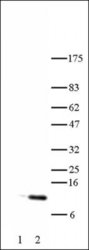
- Histone H4 acetyl Lys5 antibody tested by Western blot. HeLa acid extract probed with Histone H4 acetyl Lys5 polyclonal antibody (1:1,000 dilution). Lane 1: No treatment. Lane 2: Cells treated with sodium butyrate.

- Submitted by
- antibodies-online (provider)
- Main image

- Experimental details
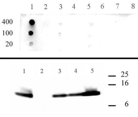
- Histone H4 acetyl Lys5 antibody specificity data. Top Panel ? Dot blot analysis was used to confirm the specificity of Histone H4 acetyl Lys5 antibody for H4 acetyl lysine 5. Peptides corresponding to regions around major sites of histone H4 acetylation were spotted onto PVDF and probed at a dilution of 1:1,000. The amount of peptide (in picomoles) spotted is indicated next to each row. Lane 1: acetyl-Lys5 peptide. Lane 2: unmodified Lys5 peptide. Lane 3: acetyl-Lys8 peptide. Lane 4: unmodified Lys8 peptide. Lane 5: acetyl-Lys12 peptide. Lane 6: unmodified Lys12 peptide. Lane 7: acetyl-Lys16 peptide. Lane 8: unmodified Lys16 peptide. Bottom Panel ? Peptide inhibition analysis. A Western blot was performed on butyrate-treated HeLa cell acid extract (5 μl per lane) at a dilution of 1:2,000. Different peptides were incubated with the antibody during the blotting procedure to determine if the recognition was blocked by a specific modification. Lane 1: no peptide. Lane 2: acetyl-Lys5 peptide. Lane 3: acetyl-Lys8 peptide. Lane 4: acetyl-Lys12 peptide. Lane 5: acetyl-Lys16 peptide.
Supportive validation
- Submitted by
- antibodies-online (provider)
- Main image
- Experimental details
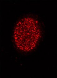
- Histone H4 acetyl Lys5 antibody tested by immunofluorescence. Staining of HeLa cells with Histone H4 acetyl Lys5 antibody (1:500 dilution, top panel) and DAPI (bottom panel).

Supportive validation
- Submitted by
- antibodies-online (provider)
- Main image
- Experimental details
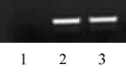
- Histone H4 acetyl Lys5 antibody tested by ChIP analysis. Chromatin IP performed on HeLa cell chromatin using 39169. PCR was performed using primers specific for the promoter region of the human GAPDH gene. Lane 1: negative IgG control. Lane 2: ChIP using 10 ul of 39169. Lane 3: Input DNA control.

- Submitted by
- antibodies-online (provider)
- Main image
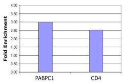
- Experimental details
- Histone H4 acetyl Lys5 antibody tested by ChIP analysis. Chromatin IP performed using the ChIP-IT® Express Kit (Catalog No. 53008) and HeLa Chromatin (1.5 x 10⁶ cell equivalents per ChIP) using 10 μl of Histone H4 acetyl Lys5 antibody or the equivalent amount of rabbit IgG as a negative control. Real time, quantitative PCR (RT-qPCR) was performed on DNA purified from each of the ChIP reactions using a primer pair specific for the indicated gene. Data are presented as Fold Enrichment of the ChIP antibody signal versus the negative control IgG using the ddCT method.
