Explore
 Validate
 Learn
Learn Western blot
Antibody data
- Antibody Data
- Antigen structure
- References [0]
- Comments [0]
- Validations
- Western blot [3]
- Immunocytochemistry [1]
Submit
Validation data
Reference
Comment
Report error
- Product number
- PA5-40698 - Provider product page

- Provider
- Invitrogen Antibodies
- Product name
- SOX11 Polyclonal Antibody
- Antibody type
- Polyclonal
- Antigen
- Synthetic peptide
- Description
- Peptide sequence: MVQQAESLEA ESNLPREALD TEEGEFMACS PVALDESDPD WCKTASGHIK
- Concentration
- 0.5 mg/mL
No comments: Submit comment
Supportive validation
- Submitted by
- Invitrogen Antibodies (provider)
- Main image
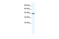
- Experimental details
- Western blot analysis of human HepG2 cell lysate using an anti-SOX11 polyclonal antibody (Product # PA5-40698).
- Submitted by
- Invitrogen Antibodies (provider)
- Main image
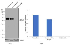
- Experimental details
- Knockdown of SOX11 was achieved by transfecting SH-SY5Y with SOX11 specific siRNAs (Silencer® select Product # s224668, s194808) . Western blot analysis (Fig. a) was performed using modified whole cell extracts (1% SDS) from the SOX11 knockdown cells (Lane 3), non-specific scrambled siRNA transfected cells (Lane 2) and untransfected cells (Lane 1). The blot was probed with SOX11 Polyclonal Antibody (Product # PA5-40698, 1:1000 dilution) and Goat anti-Rabbit IgG (H+L) Superclonal™ Recombinant Secondary Antibody, HRP conjugate (Product # A27036, 0.25µg/ml, 1:4000 dilution). Densitometric analysis of this western blot is shown i histogram (Fig. b). Loss of signal upon siRNA mediated knock down confirms that antibody is specific to SOX11.

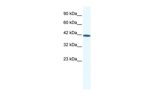
- Submitted by
- Invitrogen Antibodies (provider)
- Main image

- Experimental details
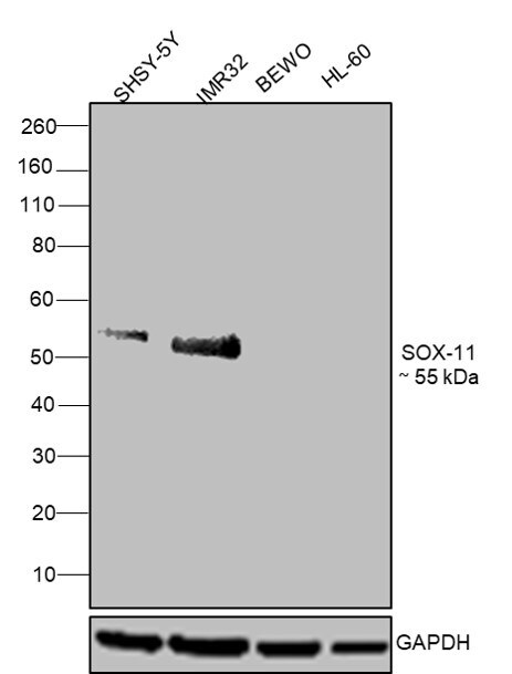
- Western blot was performed using SOX11 Polyclonal Antibody (Product # PA5-40698) and a 55 kDa band corresponding to SOX11 was observed across cell lines tested except BeWo and HL-60 which are reported to be negative. Whole cell extracts (30 µg lysate) of SHSY-5Y (Lane 1), IMR-32 (Lane 2), BeWo (Lane 3)HL-60 (Lane 4) were electrophoresed using NuPAGE® 4-12 % Bis-Tris gel (Product # NP0321BOX). Resolved proteins were then transferred onto a nitrocellulose membrane (Product # IB23001) by iBlot® 2 Dry Blotting System (Product # IB21001). The blot was probed with the primary antibody (1:1000 dilution) and detected by chemiluminescence with Goat anti-Rabbit IgG (H+L) Superclonal™ Recombinant Secondary Antibody, HRP (Product # A27036, 1:4000 dilution) using the iBright FL 1000 (Product # A32752). Chemiluminescent detection was performed using Novex® ECL Chemiluminescent Substrate Reagent Kit (Product # WP20005)..
Supportive validation
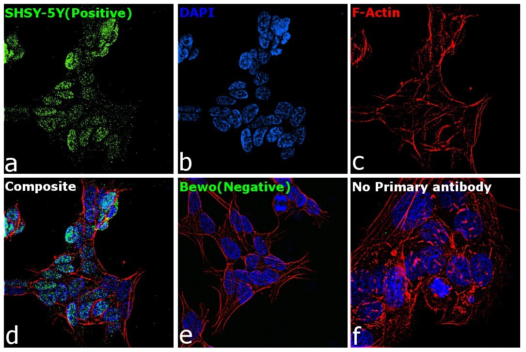
- Submitted by
- Invitrogen Antibodies (provider)
- Main image
- Experimental details
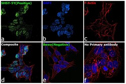
- Immunofluorescence analysis of SOX11 was performed using 70% confluent log phase SHSY-5Y and BeWo cells. The cells were fixed with 4% paraformaldehyde for 10 minutes, permeabilized with 0.1% Triton™ X-100 for 15 minutes, and blocked with 2% BSA for 1 hour at room temperature. The cells were labeled with SOX11 Polyclonal Antibody (Product # PA5-40698) at 5 µg/mL 0.1% BSA, incubated at 4 degree Celsius overnight and then labeled with Goat anti-Rabbit IgG (H+L) Superclonal™ Recombinant Secondary Antibody, Alexa Fluor® 488 conjugate (Product # A27034) at a dilution of 1:2000 for 45 minutes at room temperature (Panel a: green). Nuclei (Panel b: blue) were stained with SlowFade® Gold Antifade Mountant with DAPI (Product # S36938). F-actin (Panel c: red) was stained with Rhodamine Phalloidin (Product # R415, 1:300). Panel d represents the merged image showing localization to nucleus. Panel e shows BeWo cells with no expression of SOX11. Panel f represents control cells with no primary antibody to assess background. The images were captured at 60X magnification.