 Explore
 Validate
 Learn
Learn Western blot
Antibody data
- Antibody Data
- Antigen structure
- References [0]
- Comments [0]
- Validations
- Western blot [5]
- Immunocytochemistry [1]
- Immunohistochemistry [1]
- Chromatin Immunoprecipitation [1]
Submit
Validation data
Reference
Comment
Report error
- Product number
- PA5-47557 - Provider product page

- Provider
- Invitrogen Antibodies
- Product name
- SOX11 Polyclonal Antibody
- Antibody type
- Polyclonal
- Antigen
- Recombinant full-length protein
- Description
- In direct ELISAs, less than 5% cross-reactivity with recombinant human (rh) SOX1, rhSOX2, rhSOX3, rhSOX5, rhSOX6, rhSOX7, rhSOX9, rhSOX10, rhSOX12, rhSOX14, rhSOX15, rhSOX17, and rhSOX21 is observed.
- Concentration
- 0.2 mg/mL
No comments: Submit comment
Supportive validation
- Submitted by
- Invitrogen Antibodies (provider)
- Main image
- Experimental details
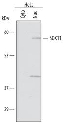
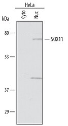
- Western blot analysis from lysates of HeLa human cervical epithelial carcinoma cell line. Gels were loaded with 25 µg of cytoplasmic (Cyto) and 25 µg of nuclear (Nuc) extracts. PVDF membrane was probed with 1 µg/mL of Sheep Anti-human SOX11 Antigen Affinity-purified Polyclonal Antibody (Product # PA5-47557) followed by HRP-conjugated Anti-Sheep IgG Secondary Antibody. A specific band was detected for SOX11 at approximately 70 kDa (as indicated). This experiment was conducted under reducing conditions.
- Submitted by
- Invitrogen Antibodies (provider)
- Main image

- Experimental details
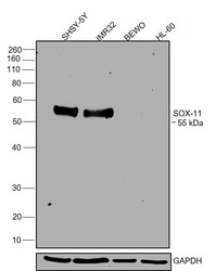
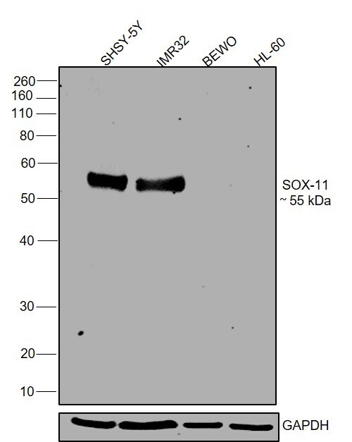
- Western blot was performed using SOX11 Polyclonal Antibody (Product # PA5-47557) and a 55 kDa band corresponding to SOX11 was observed across cell lines tested except BeWo and HL-60 which are reported to be negative. Whole cell extracts (30 µg lysate) of SHSY-5Y (Lane 1), IMR-32 (Lane 2), BeWo (Lane 3)HL-60 (Lane 4) were electrophoresed using NuPAGE® 4-12 % Bis-Tris gel (Product # NP0321BOX). Resolved proteins were then transferred onto a nitrocellulose membrane (Product # IB23001) by iBlot® 2 Dry Blotting System (Product # IB21001). The blot was probed with the primary antibody (1:1000 dilution) and detected by chemiluminescence with Rabbit anti-Sheep IgG (H+L) Secondary Antibody, HRP (Product # 61-8620, 1:4000 dilution) using the iBright FL 1000 (Product # A32752). Chemiluminescent detection was performed using Novex® ECL Chemiluminescent Substrate Reagent Kit (Product # WP20005)..

- Submitted by
- Invitrogen Antibodies (provider)
- Main image
- Experimental details
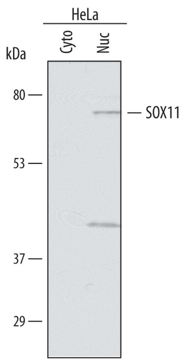
- Western blot analysis of SOX11 in HeLa human cervical epithelial carcinoma cell line. Gels were loaded with 25 µg of cytoplasmic (Cyto) and 25 µg of nuclear (Nuc) extracts. Samples were incubated in SOX11 polyclonal antibody (Product # PA5-47557) using a dilution of 1 µg/mL followed by a HRP-conjugated Anti-Sheep IgG secondary antibody. A specific band was detected for SOX11 at approximately 70 kDa (as indicated). This experiment was conducted under reducing conditions.

- Submitted by
- Invitrogen Antibodies (provider)
- Main image
- Experimental details
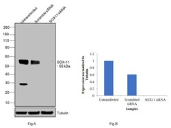
- Knockdown of SOX11 was achieved by transfecting SH-SY5Y with SOX11 specific siRNAs (Silencer® select Product # s224668, s194808). Western blot analysis (Fig. a) was performed using modified whole cell extracts (1% SDS) from the SOX11 knockdown cells (Lane 3), non-specific scrambled siRNA transfected cells (Lane 2) and untransfected cells (Lane 1). The blot was probed with SOX11 Polyclonal Antibody (Product # PA5-47557, 1:1000 dilution) and Rabbit anti-Sheep IgG (H+L) Secondary Antibody, HRP (Product # 61-8620, 1:4000 dilution). Densitometric analysis of this western blot is shown i histogram (Fig. b). Loss of signal upon siRNA mediated knock down confirms that antibody is specific to SOX11.

- Submitted by
- Invitrogen Antibodies (provider)
- Main image
- Experimental details
- Western blot was performed using SOX11 Polyclonal Antibody (Product # PA5-47557) and a 55 kDa band corresponding to SOX11 was observed across cell lines tested except BeWo and HL-60 which are reported to be negative. Whole cell extracts (30 µg lysate) of SHSY-5Y (Lane 1), IMR-32 (Lane 2), BeWo (Lane 3)HL-60 (Lane 4) were electrophoresed using NuPAGE® 4-12 % Bis-Tris gel (Product # NP0321BOX). Resolved proteins were then transferred onto a nitrocellulose membrane (Product # IB23001) by iBlot® 2 Dry Blotting System (Product # IB21001). The blot was probed with the primary antibody (1:1000 dilution) and detected by chemiluminescence with Rabbit anti-Sheep IgG (H+L) Secondary Antibody, HRP (Product # 61-8620, 1:4000 dilution) using the iBright FL 1000 (Product # A32752). Chemiluminescent detection was performed using Novex® ECL Chemiluminescent Substrate Reagent Kit (Product # WP20005)..
Supportive validation
- Submitted by
- Invitrogen Antibodies (provider)
- Main image
- Experimental details
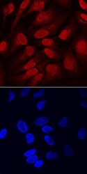
- Immunocytochemistry analysis of SOX11 in immersion fixed HeLa human cervical epithelial carcinoma cell line. Samples were incubated in SOX11 polyclonal antibody (Product # PA5-47557) using a dilution of 10 µg/mL for 3 hours at room temperature followed by NorthernLights™ 557-conjugated Anti-Sheep IgG Secondary Antibody (red, upper panel) and counterstained with DAPI (blue, lower panel). Specific staining was localized to nuclei and cytoplasm.

Supportive validation
- Submitted by
- Invitrogen Antibodies (provider)
- Main image
- Experimental details
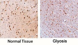
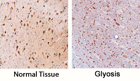
- Immunohistochemical analysis of SOX11 in immersion fixed paraffin-embedded sections of human brain (hippocampus). Samples were incubated in SOX11 polyclonal antibody (Product # PA5-47557) using a dilution of 3 µg/mL overnight at 4 °C. Tissue was stained using the Anti-Sheep HRP-DAB Cell & Tissue Staining Kit (brown) and counterstained with hematoxylin (blue). Specific staining was localized to neurons.
Supportive validation
- Submitted by
- Invitrogen Antibodies (provider)
- Main image
- Experimental details
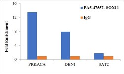
- Chromatin Immunoprecipitation (ChIP) assay of endogenous SOX11 protein using Anti-SOX11 Antibody: ChIP was performed using Anti-SOX11 Polyclonal Antibody (Product # PA5-47557) 5 µg, on sheared chromatin from SH-SY5Y cells using the MAGnify ChIP System kit (Product # 49-2024). Normal Rabbit IgG was used as a negative IP control. The purified DNA was analyzed by qPCR using primers binding to transcriptional start site of PRKACA, DBN1 and SAT2 satellite repeats. Data is presented as fold enrichment of the antibody signal versus the negative control IgG using the comparative CT method.
