Explore
 Validate
 Learn
Learn Western blot
Antibody data
- Antibody Data
- Antigen structure
- References [8]
- Comments [0]
- Validations
- Western blot [1]
- Immunocytochemistry [2]
Submit
Validation data
Reference
Comment
Report error
- Product number
- MA1-06325 - Provider product page

- Provider
- Invitrogen Antibodies
- Product name
- Cytokeratin 17 Monoclonal Antibody (E3)
- Antibody type
- Monoclonal
- Antigen
- Other
- Description
- MA1-06325 detects cytokeratin 17 in human and rat samples.
- Antibody clone number
- E3
- Concentration
- 1 mg/mL
Submitted references Impaired wound repair in adult endoglin heterozygous mice associated with lower NO bioavailability.
CD200-expressing human basal cell carcinoma cells initiate tumor growth.
Triple-negative breast cancer: immunohistochemical correlation with basaloid markers and prognostic value of survivin.
Keratin profiling in the developing human prostate. A different approach to understanding epithelial lineage.
Omics-based profiling of carcinoma of the breast and matched regional lymph node metastasis.
Identification of a subset of breast carcinomas characterized by expression of cytokeratin 15: relationship between CK15+ progenitor/amplified cells and pre-malignant lesions and invasive disease.
Basoluminal carcinoma: a new biologically and prognostically distinct entity between basal and luminal breast cancer.
Cytokeratin expression in lichen amyloidosus and macular amyloidosis.
Pérez-Gómez E, Jerkic M, Prieto M, Del Castillo G, Martín-Villar E, Letarte M, Bernabeu C, Pérez-Barriocanal F, Quintanilla M, López-Novoa JM
The Journal of investigative dermatology 2014 Jan;134(1):247-255
The Journal of investigative dermatology 2014 Jan;134(1):247-255
CD200-expressing human basal cell carcinoma cells initiate tumor growth.
Colmont CS, Benketah A, Reed SH, Hawk NV, Telford WG, Ohyama M, Udey MC, Yee CL, Vogel JC, Patel GK
Proceedings of the National Academy of Sciences of the United States of America 2013 Jan 22;110(4):1434-9
Proceedings of the National Academy of Sciences of the United States of America 2013 Jan 22;110(4):1434-9
Triple-negative breast cancer: immunohistochemical correlation with basaloid markers and prognostic value of survivin.
Dogu GG, Ozkan M, Ozturk F, Dikilitas M, Er O, Ozturk A
Medical oncology (Northwood, London, England) 2010 Mar;27(1):34-9
Medical oncology (Northwood, London, England) 2010 Mar;27(1):34-9
Keratin profiling in the developing human prostate. A different approach to understanding epithelial lineage.
Trompetter M, Smedts F, van der Wijk J, Schoots C, de Jong HJ, Hopman A, de la Rosette J
Anticancer research 2008 Jan-Feb;28(1A):237-43
Anticancer research 2008 Jan-Feb;28(1A):237-43
Omics-based profiling of carcinoma of the breast and matched regional lymph node metastasis.
Li J, Gromov P, Gromova I, Moreira JM, Timmermans-Wielenga V, Rank F, Wang K, Li S, Li H, Wiuf C, Yang H, Zhang X, Bolund L, Celis JE
Proteomics 2008 Dec;8(23-24):5038-52
Proteomics 2008 Dec;8(23-24):5038-52
Identification of a subset of breast carcinomas characterized by expression of cytokeratin 15: relationship between CK15+ progenitor/amplified cells and pre-malignant lesions and invasive disease.
Celis JE, Gromova I, Cabezón T, Gromov P, Shen T, Timmermans-Wielenga V, Rank F, Moreira JM
Molecular oncology 2007 Dec;1(3):321-49
Molecular oncology 2007 Dec;1(3):321-49
Basoluminal carcinoma: a new biologically and prognostically distinct entity between basal and luminal breast cancer.
Laakso M, Tanner M, Nilsson J, Wiklund T, Erikstein B, Kellokumpu-Lehtinen P, Malmström P, Wilking N, Bergh J, Isola J
Clinical cancer research : an official journal of the American Association for Cancer Research 2006 Jul 15;12(14 Pt 1):4185-91
Clinical cancer research : an official journal of the American Association for Cancer Research 2006 Jul 15;12(14 Pt 1):4185-91
Cytokeratin expression in lichen amyloidosus and macular amyloidosis.
Apaydin R, Gürbüz Y, Bayramgürler D, Müezzinoglu B, Bilen N
Journal of the European Academy of Dermatology and Venereology : JEADV 2004 May;18(3):305-9
Journal of the European Academy of Dermatology and Venereology : JEADV 2004 May;18(3):305-9
No comments: Submit comment
Supportive validation
- Submitted by
- Invitrogen Antibodies (provider)
- Main image

- Experimental details
- Western blot was performed using Anti- Cytokeratin 17 Monoclonal Antibody (Product # MA1-06325) and a 48 kDa band corresponding to Cytokeratin 17 was observed across cell lines tested except in MCF7 and PC-3. Whole cell extracts (30 µg lysate) of HeLa (Lane 1), A-431 (Lane 2), MCF 10A (Lane 3), MCF-7 (Lane 4), PC-3 (Lane 5) and SiHa (Lane 6) were electrophoresed using NuPAGE™ 4-12% Bis-Tris Protein Gel (Product # NP0322BOX). Resolved proteins were then transferred onto a nitrocellulose membrane (Product # IB23001) by iBlot® 2 Dry Blotting System (Product # IB21001). The blot was probed with the primary antibody (1:1000 dilution) and detected by chemiluminescence with Goat anti-Mouse IgG (H+L) Superclonal™ Recombinant Secondary Antibody, HRP (Product # A28177, 1:4000 dilution) using the iBright FL 1000 (Product # A32752). Chemiluminescent detection was performed using Novex® ECL Chemiluminescent Substrate Reagent Kit (Product # WP20005).

Supportive validation
- Submitted by
- Invitrogen Antibodies (provider)
- Main image
- Experimental details
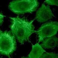
- Immunocytochemistry analysis of Cytokeratin 17 in a HaCaT keratinocyte cell culture using a Cytokeratin 17 Monoclonal Antibody (E3) (Product # MA1-06325) at a dilution of 1:500 (ice-cold methanol fixation).

- Submitted by
- Invitrogen Antibodies (provider)
- Main image
- Experimental details
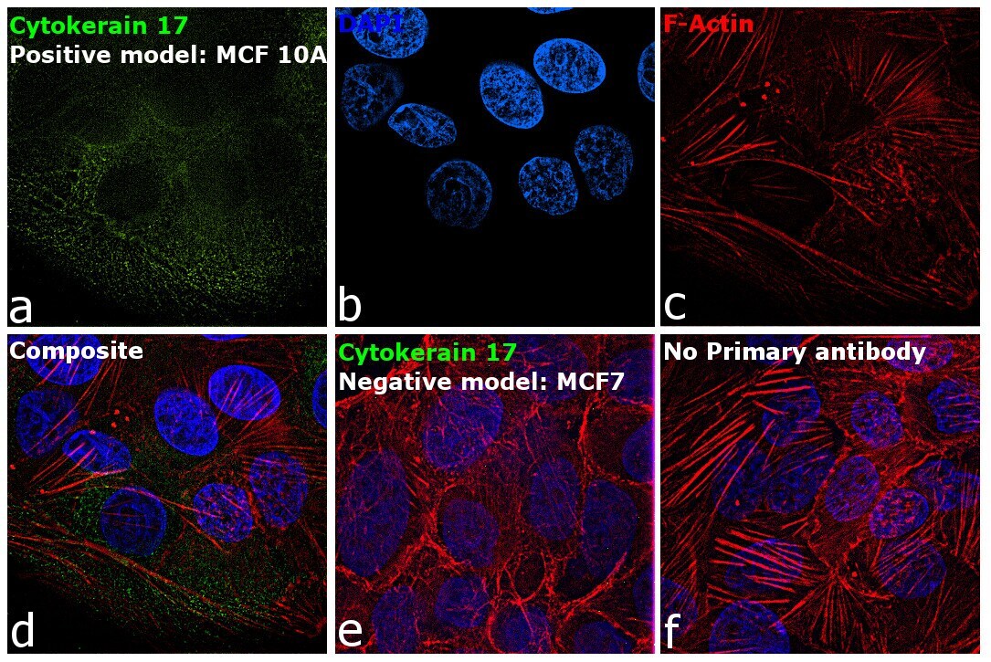
- Immunofluorescence analysis of Cytokeratin 17 was performed using MCF 10A and MCF7 cells. The cells were fixed with 4% paraformaldehyde for 10 minutes, permeabilized with 0.1% Triton™ X-100 for 15 minutes, and blocked with 2% BSA for 1 hour at room temperature. The cells were labeled with Cytokeratin 17 Monoclonal Antibody (Product # MA1-06325) at 1:100 dilution in 0.1% BSA and incubated overnight at 4 degree and then labeled with Donkey anti-Mouse IgG (H+L) Highly Cross-Adsorbed Secondary Antibody, Alexa Fluor Plus 488 conjugate (Product # A32766) at a dilution of 1:2000 for 45 minutes at room temperature (Panel a: green). Nuclei (Panel b: blue) were stained with ProLong™ Diamond Antifade Mountant with DAPI (Product # P36962). F-actin (Panel c: red) was stained with Rhodamine Phalloidin (Product # R415, 1:300). Panel d represents the composite image showing cytoskeletal localization of Keratin 17 in MCF 10A cells but not in MCF7 cells (panel e) which are reported to be low to negative for its expression. Panel f represents control cells with no primary antibody to assess background. The images were captured at 60X magnification.