 Explore
 Validate
 Learn
Learn Western blot
Antibody data
- Antibody Data
- Antigen structure
- References [14]
- Comments [0]
- Validations
- Western blot [3]
- Immunohistochemistry [1]
- Other assay [5]
Submit
Validation data
Reference
Comment
Report error
- Product number
- 14-6093-81 - Provider product page

- Provider
- Invitrogen Antibodies
- Product name
- CX3CR1 Polyclonal Antibody, eBioscience™
- Antibody type
- Polyclonal
- Antigen
- Other
- Description
- Description: The polyclonal rabbit antibody reacts with human, mouse, and rat Fractalkine receptor; the antibody was raised against a peptide corresponding to amino acids 2 to 21 of human Fractalkine receptor (CX3CR1), this sequence of human CX3CR1 differs from those of mouse and rat by four amino acids. CX3CR1 is one of the chemokine receptors that are required as coreceptors for HIV infection. The genes encoding human, murine, and rat CX3CR1 were cloned and designated V28 and CMKBRL1, CX3CR1 and RBS11, respectively. The encoded seven transmembrane protein was recently identified as the receptor for a novel transmembrane molecule, fractalkine, and renamed CX3CR1. Recently, CX3CR1 was found to serve as a coreceptor for HIV-1 and HIV-2 envelope fusion and virus infection, which can be inhibited by fractokine. CX3CR1 mediates leukocyte migration and adhesion. CX3CR1 is expressed in a variety of human tissues and cell lines.
- Concentration
- 1 mg/mL
Submitted references Modulation of inflammatory responses by fractalkine signaling in microglia.
Altered cell and RNA isoform diversity in aging Down syndrome brains.
PDTC ameliorates neuropathic pain by inhibiting microglial activation via blockage of the TNFα-CX3CR1 pathway.
Germline nuclear-predominant Pten murine model exhibits impaired social and perseverative behavior, microglial activation, and increased oxytocinergic activity.
Acute and Persistent Alterations of Cerebellar Inflammatory Networks and Glial Activation in a Rat Model of Pediatric Mild Traumatic Brain Injury.
Plasmodium falciparum proteins involved in cytoadherence of infected erythrocytes to chemokine CX3CL1.
Liver-resident NK cells confer adaptive immunity in skin-contact inflammation.
Fractalkine is expressed in the human ovary and increases progesterone biosynthesis in human luteinised granulosa cells.
Human renal cell carcinoma induces a dendritic cell subset that uses T-cell crosstalk for tumor-permissive milieu alterations.
Gene cloning, RNA distribution, and functional expression of mCX3CR1, a mouse chemotactic receptor for the CX3C chemokine fractalkine.
Identification and molecular characterization of fractalkine receptor CX3CR1, which mediates both leukocyte migration and adhesion.
The orphan G-protein-coupled receptor-encoding gene V28 is closely related to genes for chemokine receptors and is expressed in lymphoid and neural tissues.
Cloning, chromosomal localization, and RNA expression of a human beta chemokine receptor-like gene.
cDNA cloning of a G-protein-coupled receptor expressed in rat spinal cord and brain related to chemokine receptors.
Inoue K, Morimoto H, Ohgidani M, Ueki T
PloS one 2021;16(5):e0252118
PloS one 2021;16(5):e0252118
Altered cell and RNA isoform diversity in aging Down syndrome brains.
Palmer CR, Liu CS, Romanow WJ, Lee MH, Chun J
Proceedings of the National Academy of Sciences of the United States of America 2021 Nov 23;118(47)
Proceedings of the National Academy of Sciences of the United States of America 2021 Nov 23;118(47)
PDTC ameliorates neuropathic pain by inhibiting microglial activation via blockage of the TNFα-CX3CR1 pathway.
Li X, Ye Z, Guo Q, Wang E, Pan Y
European journal of histochemistry : EJH 2021 Mar 12;65(1)
European journal of histochemistry : EJH 2021 Mar 12;65(1)
Germline nuclear-predominant Pten murine model exhibits impaired social and perseverative behavior, microglial activation, and increased oxytocinergic activity.
Sarn N, Thacker S, Lee H, Eng C
Molecular autism 2021 Jun 4;12(1):41
Molecular autism 2021 Jun 4;12(1):41
Acute and Persistent Alterations of Cerebellar Inflammatory Networks and Glial Activation in a Rat Model of Pediatric Mild Traumatic Brain Injury.
Fraunberger EA, DeJesus P, Zanier ER, Shutt TE, Esser MJ
Journal of neurotrauma 2020 Jun 1;37(11):1315-1330
Journal of neurotrauma 2020 Jun 1;37(11):1315-1330
Plasmodium falciparum proteins involved in cytoadherence of infected erythrocytes to chemokine CX3CL1.
Hermand P, Cicéron L, Pionneau C, Vaquero C, Combadière C, Deterre P
Scientific reports 2016 Sep 22;6:33786
Scientific reports 2016 Sep 22;6:33786
Liver-resident NK cells confer adaptive immunity in skin-contact inflammation.
Peng H, Jiang X, Chen Y, Sojka DK, Wei H, Gao X, Sun R, Yokoyama WM, Tian Z
The Journal of clinical investigation 2013 Apr;123(4):1444-56
The Journal of clinical investigation 2013 Apr;123(4):1444-56
Fractalkine is expressed in the human ovary and increases progesterone biosynthesis in human luteinised granulosa cells.
Huang S, Zhao P, Yang L, Chen Y, Yan J, Duan E, Qiao J
Reproductive biology and endocrinology : RB&E 2011 Jun 30;9:95
Reproductive biology and endocrinology : RB&E 2011 Jun 30;9:95
Human renal cell carcinoma induces a dendritic cell subset that uses T-cell crosstalk for tumor-permissive milieu alterations.
Figel AM, Brech D, Prinz PU, Lettenmeyer UK, Eckl J, Turqueti-Neves A, Mysliwietz J, Anz D, Rieth N, Muenchmeier N, Buchner A, Porubsky S, Siegert SI, Segerer S, Nelson PJ, Noessner E
The American journal of pathology 2011 Jul;179(1):436-51
The American journal of pathology 2011 Jul;179(1):436-51
Gene cloning, RNA distribution, and functional expression of mCX3CR1, a mouse chemotactic receptor for the CX3C chemokine fractalkine.
Combadiere C, Gao J, Tiffany HL, Murphy PM
Biochemical and biophysical research communications 1998 Dec 30;253(3):728-32
Biochemical and biophysical research communications 1998 Dec 30;253(3):728-32
Identification and molecular characterization of fractalkine receptor CX3CR1, which mediates both leukocyte migration and adhesion.
Imai T, Hieshima K, Haskell C, Baba M, Nagira M, Nishimura M, Kakizaki M, Takagi S, Nomiyama H, Schall TJ, Yoshie O
Cell 1997 Nov 14;91(4):521-30
Cell 1997 Nov 14;91(4):521-30
The orphan G-protein-coupled receptor-encoding gene V28 is closely related to genes for chemokine receptors and is expressed in lymphoid and neural tissues.
Raport CJ, Schweickart VL, Eddy RL Jr, Shows TB, Gray PW
Gene 1995 Oct 3;163(2):295-9
Gene 1995 Oct 3;163(2):295-9
Cloning, chromosomal localization, and RNA expression of a human beta chemokine receptor-like gene.
Combadiere C, Ahuja SK, Murphy PM
DNA and cell biology 1995 Aug;14(8):673-80
DNA and cell biology 1995 Aug;14(8):673-80
cDNA cloning of a G-protein-coupled receptor expressed in rat spinal cord and brain related to chemokine receptors.
Harrison JK, Barber CM, Lynch KR
Neuroscience letters 1994 Mar 14;169(1-2):85-9
Neuroscience letters 1994 Mar 14;169(1-2):85-9
No comments: Submit comment
Supportive validation
- Submitted by
- Invitrogen Antibodies (provider)
- Main image

- Experimental details
- Immunoblot analysis of reduced human spleen lysates using Anti-CX3CR1 Purified and detected using Anti-Rabbit IgG-HRP.

- Submitted by
- Invitrogen Antibodies (provider)
- Main image

- Experimental details
- Knockdown of CX3CR1 was achieved by transfecting HeLa with CX3CR1 specific siRNAs (Silencer® select Product # s3772, s3771). Western blot analysis (Fig. a) was performed using membrane enriched extracts from the CX3CR1 knockdown cells (lane 3), non-specific scrambled siRNA transfected cells (lane 2) and untransfected cells (lane 1). The blot was probed with CX3CR1 Polyclonal Antibody, eBioscience (Product # 14-6093-81, 1:1000 dilution) and Goat anti-Rabbit IgG (H+L) Superclonal™ Recombinant Secondary Antibody, HRP (Product # A27036, 1:4000 dilution). Densitometric analysis of this western blot is shown in histogram (Fig. b). Decrease in CX3CR1 signal was observed in siRNA transfected lysates.

- Submitted by
- Invitrogen Antibodies (provider)
- Main image

- Experimental details
- Western blot was performed using Anti-CX3CR1 Polyclonal Antibody (Product # 14-6093-81) and a 52 kDa band corresponding to CX3CR1 was observed in THP-1, SH-SY5Y, MDA-MB-231, NK-92, SK-O-V3, HeLa, Caco-2 and Hep G2. Membrane enriched extracts (30 µg lysate) of THP-1 (Lane 1),SH-SY5Y (Lane 2), MDA-MB-231 (Lane 3), NK-92 (Lane 4), SK-O-V3 (Lane 5), HeLa (Lane 6), Caco-2 (Lane 7) and Hep G2 (Lane 8) were electrophoresed using Novex® NuPAGE® 4-12 % Bis-Tris gel (Product # NP0321BOX). Resolved proteins were then transferred onto a nitrocellulose membrane (Product # IB23001) by iBlot® 2 Dry Blotting System (Product # IB21001). The blot was probed with the primary antibody (1:1,000 dilution) and detected by chemiluminescence with Goat anti-Rabbit IgG (H+L) Superclonal™ Recombinant Secondary Antibody, HRP (Product # A27036, 1:4000 dilution). Chemiluminescent detection was performed using Novex® ECL Chemiluminescent Substrate Reagent Kit (Product # WP20005). * - There was an uncharacterized band observed in THP-1 and SK-O-V3 at 100kDa.

Supportive validation
- Submitted by
- Invitrogen Antibodies (provider)
- Main image

- Experimental details
- Immunohistochemical staining with antigen retreival of formalin-fixed, paraffin-embedded human heart tissue using Anti-CX3CR1 Purified at 2 µg/mL and detected using Anti-Rabbit IgG-HRP.

Supportive validation
- Submitted by
- Invitrogen Antibodies (provider)
- Main image
- Experimental details
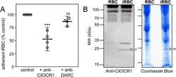
- Figure 2 Electrophoresis and anti-CX3CR1 staining of 3D7-iRBC membranes. ( A ) Static adherence to CX3CL1 of enriched 3D7-iRBC pretreated or not (control) with 0.5 mug/ml of anti-CX3CR1 or anti-DARC antibodies, in 96-well plates coated with 25 pmoles. The number of adherent cells was expressed as percent of the control, as mean values and standard deviations from four independent experiments. ANOVA followed by post hoc analysis with Tukey test was performed to establish the levels of significance: ***p 0.05. ( B ) Membranes proteins of normal RBC and 3D7-iRBC were fractionated on SDS/PAGE and transferred to PVDF membrane, immunostained with anti-CX3CR1 (1/500 dilution, left panel) or stained with Coomassie blue (right panel). Arrows indicates the gel fraction used for mass spectrometry analysis. Arrows indicates the gel fraction used for mass spectrometry analysis. The 20-25 kDa band was the only band specifically appearing in the iRBC membrane lysates (left panel).

- Submitted by
- Invitrogen Antibodies (provider)
- Main image

- Experimental details
- Figure 6. Effect of PDTC on CX3CR1 protein expression in TNFalpha-induced microglial cells. PDTC-pre-treatment group had lower CX3CR1 protein expression than other groups did. A) Bands of western blotting of CX3CR1 protein expression. B) Statistical analysis of relative density of western blotting bands is shown between different groups. Results are expressed as mean+- SEM (n=4). #p

- Submitted by
- Invitrogen Antibodies (provider)
- Main image

- Experimental details
- Fig. 4 In vitro validation of Pten Y68H microglia pathology. a Representative immunofluorescence staining of Pten + / + and Pten Y68H/ + primary microglia with Pten (red), Iba1 (red), C1q (green), Trem2 (green), Cx3cr1 (green), and DAPI (blue). N = 7. b-e Quantification of IF staining in panel a for Pten ( P = 0.0006; b) , Iba1 ( P = 0.038; c ), C1q ( P = 0.004; d ), Trem2 ( P = NS; e ), and Cx3cr1 ( P = NS; f ). g Representative image of phagocytosis assay comparing Pten + / + and Pten Y68H/ + microglia. h Quantification of panel g for proportion of active phagocytic microglia over total microglia. P = 0.01 i Quantification of panel g of average bead counts per phagocytic microglia. P = 0.005. P value key: * P < 0.05, ** P < 0.01, *** P < 0.001

- Submitted by
- Invitrogen Antibodies (provider)
- Main image

- Experimental details
- 10.1371/journal.pone.0252118.g003 Fig 3 CX3CR1 overexpression attenuates LPS-induced inflammatory responses. (A) CX3CR1-HA-expressing or mock plasmids were transfected into the BV-2 cells. The cells were collected 2 days later, and the RNAs were extracted, followed by cDNA synthesis and quantitative real-time PCR. n = 3. The images show the detection of exogenous CX3CR1 using antibodies for CX3CR1 and HA. An HA-tagged taurine transporter was used as a negative control. The cells were stained with DAPI. Scale bar = 20 mum. (B) CX3CR1-HA-expressing or mock plasmids were transfected into the BV-2 cells. After 2 days, the cells were treated with LPS, and FKN was administered 4 h later. Twenty hours later, the media were collected, and the NO concentration was measured. n = 3-4. ** p < 0.01 vs. mock, ## p < 0.01 vs. control, two-way ANOVA followed by Sidak's multiple comparison test. (C) The cells were either untreated or treated with LPS for 24 h. The media were collected, and FKN was measured using ELISA. n = 3-4. * p < 0.05 vs. LPS -, Student's t -test. (D) A CX3CR1-HA-expressing plasmid or a mock vector was transfected into the BV-2 cells. Two days later, they were treated with LPS for 4 h, and the RNAs were extracted, followed by cDNA synthesis and quantitative real-time PCR. n = 3.

- Submitted by
- Invitrogen Antibodies (provider)
- Main image

- Experimental details
- Fig. 4. Hallmarks of microglial activation in DS microglia. ( A ) UMAP of microglia from all processed samples, colored by microglial subcluster. ( B ) Heatmap displaying key differentially expressed genes used to define microglial subclusters. ( C ) Fraction of total microglia from each sample that clustered in each of the four major microglia subclusters. ( D ) Violin plots of gene expression for hallmark microglial activation genes from DS vs. Ctrl and DS-young vs. Ctrl-young cohorts; adjusted P value using Bonferroni correction on Wilcoxon rank sum test. ( E ) Western blot for CX3CR1 and quantification relative to GAPDH. Asterisk denotes statistical significance in unpaired t test ( P = 0.011). ( F ) Violin plots of gene expression for C1q complement genes, ADGRG1 , and RUNX1 . ( G ) Volcano plots for total DEGs in microglia (gray), DEGs from HSA21 that are triplicated in the Dp16 mouse model but not Tc1 or Ts1Rhr (green), and all other HSA21 microglial DEGs (purple) (-).
