Explore
 Validate
 Learn
Learn Western blot
 Immunocytochemistry
Antibody data
- Antibody Data
- Antigen structure
- References [0]
- Comments [0]
- Validations
- Western blot [2]
- Immunohistochemistry [1]
Submit
Validation data
Reference
Comment
Report error
- Product number
- NBP2-54790 - Provider product page

- Provider
- Novus Biologicals
- Product name
- Rabbit Monoclonal CD36/SR-B3 Antibody
- Antibody type
- Monoclonal
- Description
- Protein A or G purified.
- Reactivity
- Human
- Host
- Rabbit
- Isotype
- IgG
- Vial size
- 0.1 mg
- Concentration
- 1.0 mg/ml
- Storage
- Store at 4C short term. Aliquot and store at -20C long term. Avoid freeze-thaw cycles.
No comments: Submit comment
Supportive validation
- Submitted by
- Novus Biologicals (provider)
- Main image
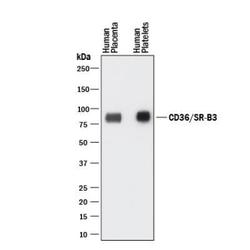
- Experimental details
- Western Blot: CD36/SR-B3 Antibody (1283D) [NBP2-54790] - Western blot shows lysates of human placenta tissue and human platelets. PVDF membrane was probed with 1 ug/ml of Rabbit Anti-Human CD36/SR-B3 Monoclonal antibody (catalog number NBP2-54790) followed by HRP-conjugated Anti-Rabbit IgG Secondary Antibody (catalog number HAF008). A specific band was detected for CD36/SR-B3 at approximately 85 kDa (as indicated). This experiment was conducted under reducing conditions.

- Submitted by
- Novus Biologicals (provider)
- Main image

- Experimental details
- Western Blot: CD36/SR-B3 Antibody (1283D) [NBP2-54790] - Total protein from human skin and adipose tissue was separated on a 12% gel by SDS-PAGE, transferred to PVDF membrane and blocked in 5% non-fat milk in TBST. The membrane was probed with 1.0 ug/ml anti-CD36 in 1% non-fat milk in TBST and detected with an anti-rabbit HRP secondary antibody using chemiluminescence.

Supportive validation
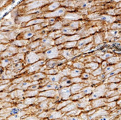
- Submitted by
- Novus Biologicals (provider)
- Main image
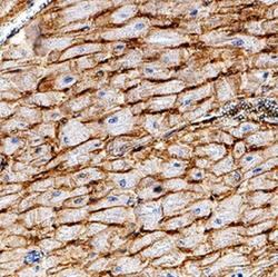
- Experimental details
- Immunohistochemistry-Paraffin: CD36/SR-B3 Antibody (1283D) [NBP2-54790] - CD36/SR-B3 was detected in immersion fixed paraffin-embedded sections of human heart using Rabbit Anti-Human CD36/SR-B3 Monoclonal Antibody for 1 hour at room temperature followed by incubation with the Anti-Rabbit IgG VisUCyte™ HRP Polymer Antibody. Tissue was stained using DAB (brown) and counterstained with hematoxylin (blue). Specific staining was localized to cardiomyocyte membranes.