 Explore
 Validate
 Learn
Learn Flow cytometry
Antibody data
- Antibody Data
- Antigen structure
- References [2]
- Comments [0]
- Validations
- Flow cytometry [1]
Submit
Validation data
Reference
Comment
Report error
- Product number
- 48-9033-41 - Provider product page

- Provider
- Invitrogen Antibodies
- Product name
- Anti-Phospho-STAT3 (Tyr705) Monoclonal Antibody (LUVNKLA), eFluor 450, eBioscience™
- Antibody type
- Monoclonal
- Antigen
- Other
- Description
- Description: This LUVNKLA monoclonal antibody recognizes human and mouse signal transducer and activator of transcription 3 (STAT3) when phosphorylated on tyrosine 705 (Y705). The STAT family represents seven transcription factors (STATs 1, 2, 3, 4, 5A, 5B, and 6) that are involved in many cellular processes including apoptosis, cell differentiation, and proliferation in a cell type- and cytokine-specific manner. STAT proteins are activated by ligand binding to cytokine receptors that associate with Janus kinase (JAK) family members. Following their phosphorylation by JAKs, STAT proteins translocate to the nucleus where they bind to DNA and regulate transcription of specific genes in a cell type- and cytokine-specific manner. STAT3 is activated downstream of numerous cytokines including interferons, IL-5, IL-6, IL-10, and LIF. STAT3 is important for the differentiation of Th17 cells and mediates a variety of cellular processes including cell growth and survival. The importance of STAT3 is highlighted by both loss-of-function and gain-of-function mutations. Deletion of STAT3 in T cells results in decreased IL-6- and IL-2-mediated proliferation, while deletion of STAT3 in neutrophils and macrophages results in increased susceptibility to LPS-induced endotoxic shock and increased production of the pro-inflammatory cytokines IL-6 and TNF alpha. Hyper STAT3 activity is associated with poor prognosis of many different cancers. Applications Reported: This LUVNKLA antibody has been reported for use in intracellular staining followed by flow cytometric analysis. Applications Tested: This LUVNKLA antibody has been pre-titrated and tested by intracellular staining followed by flow cytometric analysis of stimulated normal human peripheral blood cells. This can be used at 5 µL (0.06 µg) per test. A test is defined as the amount (µg) of antibody that will stain a cell sample in a final volume of 100 µL. Cell number should be determined empirically but can range from 10^5 to 10^8 cells/test. Staining Protocol: We recommend using Protocol C: Two-step protocol: Fixation/Methanol. Protocol A: Two-step protocol: intracellular (cytoplasmic) proteins and Protocol B: One-step protocol: intracellular (nuclear) proteins cannot be used. All Protocols can be found in the Flow Cytometry Protocols: "Staining Intracellular Antigens for Flow Cytometry Protocol" located in the Best Protocols Section under the Resources tab online. eFluor® 450 is an alternative to Pacific Blue®. eFluor® 450 emits at 445 nm and is excited with the Violet laser (405 nm). Please make sure that your instrument is capable of detecting this fluorochome. Excitation: 405 nm; Emission: 445 nm; Laser: Violet Laser. Filtration: 0.2 µm post-manufacturing filtered.
- Reactivity
- Human, Mouse
- Host
- Mouse
- Isotype
- IgG
- Antibody clone number
- LUVNKLA
- Vial size
- 25 Tests
- Concentration
- 5 µL/Test
- Storage
- 4° C, store in dark, DO NOT FREEZE!
Submitted references ATF3 Sustains IL-22-Induced STAT3 Phosphorylation to Maintain Mucosal Immunity Through Inhibiting Phosphatases.
Receptor-mediated dimerization of JAK2 FERM domains is required for JAK2 activation.
Glal D, Sudhakar JN, Lu HH, Liu MC, Chiang HY, Liu YC, Cheng CF, Shui JW
Frontiers in immunology 2018;9:2522
Frontiers in immunology 2018;9:2522
Receptor-mediated dimerization of JAK2 FERM domains is required for JAK2 activation.
Ferrao RD, Wallweber HJ, Lupardus PJ
eLife 2018 Jul 25;7
eLife 2018 Jul 25;7
No comments: Submit comment
Supportive validation
- Submitted by
- Invitrogen Antibodies (provider)
- Main image
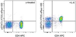
- Experimental details
- Intracellular staining of untreated (left) or 15-minute IL-6 treated (right) normal human peripheral blood cells with Anti-Human CD4 APC (Product # 17-0049-42) and Anti-Human/Mouse phospho-STAT3 (Y705) eFluor® 450. Cells in the lymphocyte gate were used for analysis. Cells were stained using the Intracellular Fixation/Methanol protocol.
