 Explore
 Validate
 Learn
Learn Western blot
 ELISA
ELISAAntibody data
- Antibody Data
- Antigen structure
- References [2]
- Comments [0]
- Validations
- Western blot [4]
- Immunocytochemistry [1]
Submit
Validation data
Reference
Comment
Report error
- Product number
- 33-2300 - Provider product page

- Provider
- Invitrogen Antibodies
- Product name
- STAT4 Monoclonal Antibody (ST4-5D6)
- Antibody type
- Monoclonal
- Antigen
- Synthetic peptide
- Reactivity
- Human, Mouse, Rat
- Host
- Mouse
- Isotype
- IgG
- Antibody clone number
- ST4-5D6
- Vial size
- 100 µg
- Concentration
- 0.5 mg/mL
- Storage
- -20°C
Submitted references Increased sensitivity to interferon-alpha in psoriatic T cells.
Activated signal transducer and activator of transcription (STAT) 3: localization in focal adhesions and function in ovarian cancer cell motility.
Eriksen KW, Lovato P, Skov L, Krejsgaard T, Kaltoft K, Geisler C, Ødum N
The Journal of investigative dermatology 2005 Nov;125(5):936-44
The Journal of investigative dermatology 2005 Nov;125(5):936-44
Activated signal transducer and activator of transcription (STAT) 3: localization in focal adhesions and function in ovarian cancer cell motility.
Silver DL, Naora H, Liu J, Cheng W, Montell DJ
Cancer research 2004 May 15;64(10):3550-8
Cancer research 2004 May 15;64(10):3550-8
No comments: Submit comment
Supportive validation
- Submitted by
- Invitrogen Antibodies (provider)
- Main image
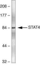
- Experimental details
- Western blot analysis of mouse testis homogenates using Ms anti-STAT4 (Product # 33-2300)

- Submitted by
- Invitrogen Antibodies (provider)
- Main image

- Experimental details
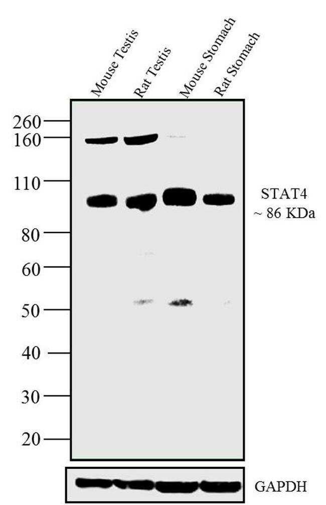
- Western blot analysis was performed on whole cell extracts (30 µg lysate) of Mouse testis (Lane 1), Rat testis (Lane 2), Mouse Stomach (Lane 3), Rat stomach (Lane 4). The blots were probed with Anti-STAT4 Mouse Monoclonal Antibody (Product # 33-2300, 1-3 µg/mL) and detected by chemiluminescence using Goat anti-Mouse IgG (H+L) Secondary Antibody, HRP conjugate (Product # 62-6520, 1:4000 dilution). A 86 kDa band corresponding to STAT4 was observed across tissue tested along with a 160 kDa extra band in Mouse Testis and Rat Testis. Known quantity of protein samples were electrophoresed using Novex® NuPAGE® 4-12 % Bis-Tris gel (Product # NP0321BOX), XCell SureLock™ Electrophoresis System (Product # EI0002) and Novex® Sharp Pre-Stained Protein Standard (Product # LC5800). Resolved proteins were then transferred onto a transferred onto a nitrocellulose membrane with Pierce™ Power Blotter System (22834). The membrane was probed with the relevant primary and secondary Antibody following blocking with 5 % skimmed milk. Chemiluminescent detection was performed using Pierce™ ECL Western Blotting Substrate (Product # 32106).
- Submitted by
- Invitrogen Antibodies (provider)
- Main image
- Experimental details
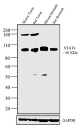
- Western blot analysis was performed on whole cell extracts (30 µg lysate) of Mouse testis (Lane 1), Rat testis (Lane 2), Mouse Stomach (Lane 3), Rat stomach (Lane 4). The blots were probed with Anti-STAT4 Mouse Monoclonal Antibody (Product # 33-2300, 1-3 µg/mL) and detected by chemiluminescence using Goat anti-Mouse IgG (H+L) Secondary Antibody, HRP conjugate (Product # 62-6520, 1:4000 dilution). A 86 kDa band corresponding to STAT4 was observed across tissue tested along with a 160 kDa extra band in Mouse Testis and Rat Testis. Known quantity of protein samples were electrophoresed using Novex® NuPAGE® 4-12 % Bis-Tris gel (Product # NP0321BOX), XCell SureLock™ Electrophoresis System (Product # EI0002) and Novex® Sharp Pre-Stained Protein Standard (Product # LC5800). Resolved proteins were then transferred onto a transferred onto a nitrocellulose membrane with Pierce™ Power Blotter System (22834). The membrane was probed with the relevant primary and secondary Antibody following blocking with 5 % skimmed milk. Chemiluminescent detection was performed using Pierce™ ECL Western Blotting Substrate (Product # 32106).

- Submitted by
- Invitrogen Antibodies (provider)
- Main image
- Experimental details
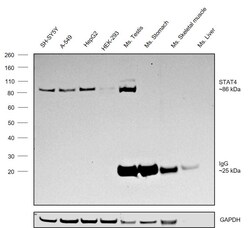
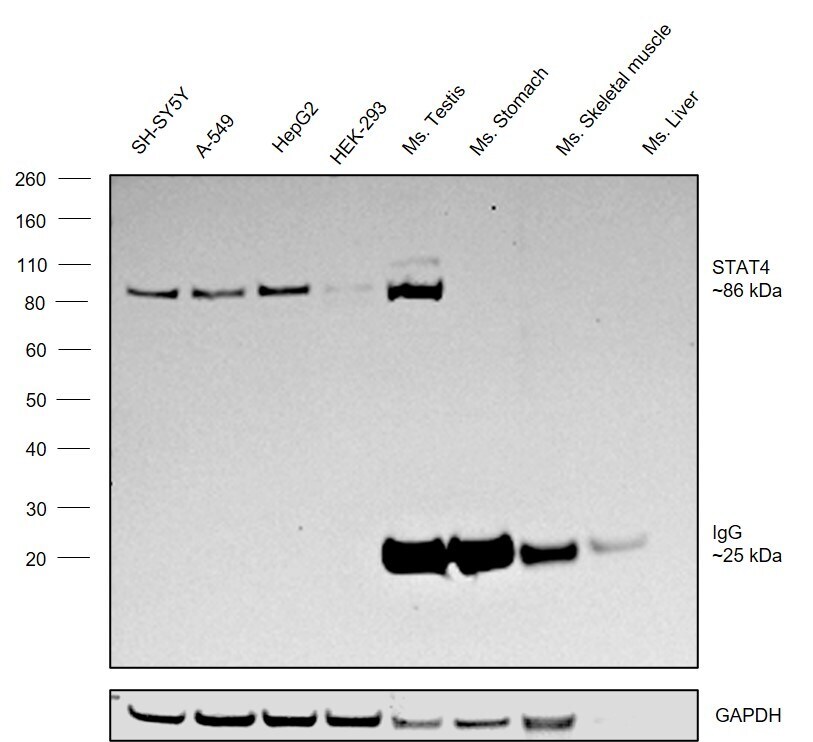
- Western blot was performed using Anti-STAT4 Monoclonal Antibody (ST4-5D6) (Product # 33-2300) and a ~86 kDa band corresponding to Signal transducer and activator of transcription 4 was observed across cell lines and tissues tested. Whole cell extracts (50 µg lysate) of SH-SY5Y (Lane 1), A549 (Lane 2), HepG2 (Lane 3), HEK-293 (Lane 4), Mouse Testis (Lane 5), Mouse Stomach (Lane 6), Mouse Skeletal Muscle (Lane 7), Mouse Liver (Lane 8) were electrophoresed using NuPAGE™ 4-12% Bis-Tris Protein Gel (Product # NP0322BOX). Resolved proteins were then transferred onto a nitrocellulose membrane (Product # IB23001) by iBlot® 2 Dry Blotting System (Product # IB21001). The blot was probed with the primary antibody (0.5 µg/mL) and detected by chemiluminescence with Goat anti-Mouse IgG (H+L) Superclonal™ Recombinant Secondary Antibody, HRP (Product # A28177,1:20000) using the iBright FL 1000 (Product # A32752). Chemiluminescent detection was performed using SuperSignal™ West Pico PLUS Chemiluminescent Substrate (Product # 34580).A 25 kDa band corresponding to IgG was observed in the mouse tissue lysates.
Supportive validation
- Submitted by
- Invitrogen Antibodies (provider)
- Main image

- Experimental details
- Immunofluorescence analysis of Signal transducer and activator of transcription 4 was performed using 70% confluent log phase SH-SY5Y cells. The cells were fixed with 4% paraformaldehyde for 10 minutes, permeabilized with 0.1% Triton™ X-100 for 15 minutes, and blocked with 2% BSA for 45 minutes at room temperature. The cells were labeled with STAT4 Monoclonal Antibody (ST4-5D6) (Product # 33-2300) at 1:100 in 0.1% BSA, incubated at 4 degree celsius overnight and then labeled with Donkey anti-Mouse IgG (H+L) Highly Cross-Adsorbed Secondary Antibody, Alexa Fluor Plus 488 (Product # A32766), (1:2000), for 45 minutes at room temperature (Panel a: Green). Nuclei (Panel b:Blue) were stained with ProLong™ Diamond Antifade Mountant with DAPI (Product # P36962). F-actin (Panel c: Red) was stained with Rhodamine Phalloidin (Product # R415, 1:300). Panel d represents the merged image showing Nuclear localization. Panel e represents control cells with no primary antibody to assess background. The images were captured at 60X magnification.
