 Explore
 Validate
 Learn
Learn Flow cytometry
Antibody data
- Antibody Data
- Antigen structure
- References [1]
- Comments [0]
- Validations
- Flow cytometry [1]
Submit
Validation data
Reference
Comment
Report error
- Product number
- 48-9008-41 - Provider product page

- Provider
- Invitrogen Antibodies
- Product name
- Anti-Phospho-STAT1 (Tyr701) Monoclonal Antibody (KIKSI0803), eFluor 450, eBioscience™
- Antibody type
- Monoclonal
- Antigen
- Other
- Description
- Description: This KIKSI0803 monoclonal antibody recognizes signal transducer and activator of transcription 1 (STAT1) when phosphorylated on tyrosine 701. STAT proteins are activated by ligand binding to receptors, such as cytokine receptors, that associate with Janus kinase (JAK) family members. Following their phosphorylation by JAKs, STAT proteins translocate to the nucleus where they bind to DNA and regulate transcription of specific genes in a cell type- and cytokine-specific manner. Phosphorylation of STAT1 on tyrosine 701 by JAK1 and JAK2 is essential for STAT1 dimer formation, nuclear translocation, and DNA binding activity. In response to IFN gamma stimulation, STAT1 homodimerizes or forms heterodimers with STAT3 that can bind to GAS (IFN gamma-activated sequence) promoter elements. In response to either IFN alpha or IFN beta stimulation, STAT1 forms a heterodimer with STAT2 that can bind ISRE (IFN-stimulated response element) promoter elements. Specificity of this KIKSI0803 clone was determined by ELISA, flow cytometry, and western blotting. Applications Reported: This KIKSI0803 antibody has been reported for use in intracellular staining followed by flow cytometric analysis. Applications Tested: This KIKSI0803 antibody has been pre-titrated and tested by intracellular staining followed by flow cytometric analysis of stimulated normal human peripheral blood cells. This can be used at 5 µL (0.125 µg) per test. A test is defined as the amount (µg) of antibody that will stain a cell sample in a final volume of 100 µL. Cell number should be determined empirically but can range from 10^5 to 10^8 cells/test. Staining Protocol: We recommend using Protocol C: Two-step protocol: Fixation/Methanol. Protocol A: Two-step protocol: intracellular (cytoplasmic) proteins and Protocol B: One-step protocol: intracellular (nuclear) proteins cannot be used. All Protocols can be found in the Flow Cytometry Protocols: "Staining Intracellular Antigens for Flow Cytometry Protocol" located in the Best Protocols Section under the Resources tab online. eFluor® 450 is an alternative to Pacific Blue®. eFluor® 450 emits at 445 nm and is excited with the Violet laser (405 nm). Please make sure that your instrument is capable of detecting this fluorochome. Excitation: 405 nm; Emission: 445 nm; Laser: Violet Laser. Filtration: 0.2 µm post-manufacturing filtered.
- Reactivity
- Human
- Host
- Mouse
- Isotype
- IgG
- Antibody clone number
- KIKSI0803
- Vial size
- 25 Tests
- Concentration
- 5 µL/Test
- Storage
- 4° C, store in dark, DO NOT FREEZE!
Submitted references Immune-Mediated Inflammation May Contribute to the Pathogenesis of Cardiovascular Disease in Mucopolysaccharidosis Type I.
Khalid O, Vera MU, Gordts PL, Ellinwood NM, Schwartz PH, Dickson PI, Esko JD, Wang RY
PloS one 2016;11(3):e0150850
PloS one 2016;11(3):e0150850
No comments: Submit comment
Supportive validation
- Submitted by
- Invitrogen Antibodies (provider)
- Main image
- Experimental details
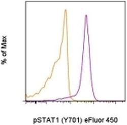
- Intracellular staining of unstimulated (orange histogram) or 15-minute IFN gamma-stimulated (purple histogram) normal human peripheral blood cells with Anti-Human phospho-STAT1 (Y701) eFluor® 450 using the Fixation/Methanol Protocol. CD4-low monocytes were used for analysis.
