Explore
 Validate
 Learn
Learn Western blot
Antibody data
- Antibody Data
- Antigen structure
- References [4]
- Comments [0]
- Validations
- Western blot [1]
Submit
Validation data
Reference
Comment
Report error
- Product number
- MA1-82934 - Provider product page

- Provider
- Invitrogen Antibodies
- Product name
- Complement C3b Monoclonal Antibody (H11)
- Antibody type
- Monoclonal
- Antigen
- Other
- Description
- Reconstitute with 1 mL of distilled water. After reconstitution, store product undiluted at 4ºC or -20ºC if preferred.
- Antibody clone number
- H11
- Concentration
- 50 µg/mL
Submitted references Overexpression of cortactin increases invasion potential in oral squamous cell carcinoma.
Combinatorial efficacy achieved through two-point blockade within a signaling pathway-a chemical genetic approach.
Physical interaction between epidermal growth factor receptor and DNA-dependent protein kinase in mammalian cells.
Physical interaction between epidermal growth factor receptor and DNA-dependent protein kinase in mammalian cells.
Yamada S, Yanamoto S, Kawasaki G, Mizuno A, Nemoto TK
Pathology oncology research : POR 2010 Dec;16(4):523-31
Pathology oncology research : POR 2010 Dec;16(4):523-31
Combinatorial efficacy achieved through two-point blockade within a signaling pathway-a chemical genetic approach.
Fan QW, Specht KM, Zhang C, Goldenberg DD, Shokat KM, Weiss WA
Cancer research 2003 Dec 15;63(24):8930-8
Cancer research 2003 Dec 15;63(24):8930-8
Physical interaction between epidermal growth factor receptor and DNA-dependent protein kinase in mammalian cells.
Bandyopadhyay D, Mandal M, Adam L, Mendelsohn J, Kumar R
The Journal of biological chemistry 1998 Jan 16;273(3):1568-73
The Journal of biological chemistry 1998 Jan 16;273(3):1568-73
Physical interaction between epidermal growth factor receptor and DNA-dependent protein kinase in mammalian cells.
Bandyopadhyay D, Mandal M, Adam L, Mendelsohn J, Kumar R
The Journal of biological chemistry 1998 Jan 16;273(3):1568-73
The Journal of biological chemistry 1998 Jan 16;273(3):1568-73
No comments: Submit comment
Supportive validation
- Submitted by
- Invitrogen Antibodies (provider)
- Main image
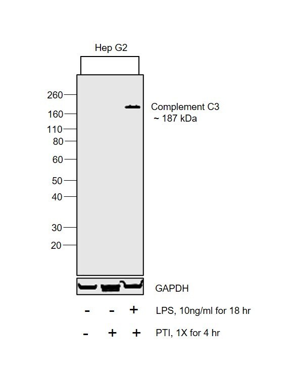
- Experimental details
- Western blot was performed using Anti-Complement C3b Monoclonal Antibody (H11) (Product # MA1-82934) and a 187 kDa band corresponding to Complement C3 was observed in HepG2 treated with LPS and PTI. Whole cell extracts (30 µg lysate) of Hep G2 (Lane 1), Hep G2 treated with protein transport inhibitor (1X for 4 hr) (Lane 2) and Hep G2 treated with LPS (10 ng/mL for 18 hr) followed by protein transport inhibitor (1X for 4 hr) (Lane 3) were electrophoresed using NuPAGE™ 4-12% Bis-Tris Protein Gel (Product # NP0322BOX). Resolved proteins were then transferred onto a Nitrocellulose membrane (Product # IB23001) by iBlot® 2 Dry Blotting System (Product # IB21001). The blot was probed with the primary antibody (1 µg/mL) and detected by chemiluminescence with Goat anti-Mouse IgG (H+L) Superclonal™ Recombinant Secondary Antibody, HRP (Product # A28177, 1:4000 dilution) using the iBright FL 1000 (Product # A32752). Chemiluminescent detection was performed using Novex® ECL Reagent Kit (Product # WP20005).