 Explore
 Validate
 Learn
Learn Western blot
Antibody data
- Antibody Data
- Antigen structure
- References [0]
- Comments [0]
- Validations
- Western blot [1]
- Immunocytochemistry [3]
- Immunohistochemistry [1]
Submit
Validation data
Reference
Comment
Report error
- Product number
- PA5-42722 - Provider product page

- Provider
- Invitrogen Antibodies
- Product name
- PTPN2 Polyclonal Antibody
- Antibody type
- Polyclonal
- Antigen
- Synthetic peptide
- Description
- Peptide sequence: ESGSLNPDHG PAVIHCSAGI GRSGTFSLVD TCLVLMEKGD DINIKQVLLN
- Concentration
- 0.5 mg/mL
No comments: Submit comment
Supportive validation
- Submitted by
- Invitrogen Antibodies (provider)
- Main image
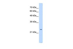
- Experimental details
- Western blot analysis of transfected 293T cells using an anti-PTPN2 / TCPTP polyclonal antibody (Product # PA5-42722).

Supportive validation
- Submitted by
- Invitrogen Antibodies (provider)
- Main image

- Experimental details
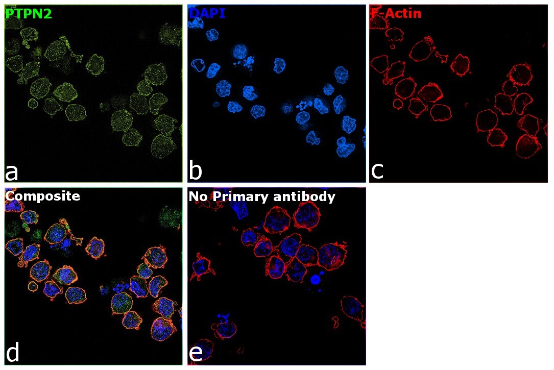
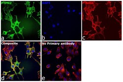
- Immunofluorescence analysis of PTPN2 (Tyrosine-protein phosphatase non-receptor type 2) was performed using K-562 cells. The cells were fixed with 4% paraformaldehyde for 10 minutes, permeabilized with 0.1% Triton™ X-100 for 10 minutes, and blocked with 2% BSA for 45 minutes at room temperature. The cells were labeled with PTPN2 Polyclonal Antibody (Product # PA5-42722) at 1:100 in 0.1% BSA, incubated at 4 degree celsius overnight and then labeled with Donkey anti-Rabbit IgG (H+L) Highly Cross-Adsorbed Secondary Antibody, Alexa Fluor Plus 488 (Product # A32790), (1:2500 dilution), for 45 minutes at room temperature (Panel a: Green). Nuclei (Panel b:Blue) were stained with ProLong™ Diamond Antifade Mountant with DAPI (Product # P36962). F-actin (Panel c: Red) was stained with Rhodamine Phalloidin (Product # R415, 1:300). Panel d represents the merged image showing cytoplasmic localization of PTPN2 in K-562 cells. Panel e represents control cells with no primary antibody to assess background. The images were captured at 60X magnification.
- Submitted by
- Invitrogen Antibodies (provider)
- Main image
- Experimental details
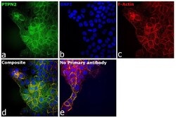
- Immunofluorescence analysis of PTPN2 was performed using 70% confluent log phase A-431 cells. The cells were fixed with 4% paraformaldehyde for 10 minutes, permeabilized with 0.01% Triton™ X-100 for 15 minutes, and blocked with 2% BSA for 1 hour at room temperature. The cells were labeled with PTPN2 Polyclonal Antibody (Product # PA5-42722) at 1:100 dilution in 0.1% BSA, incubated at 4 degree celsius overnight and then with Goat anti-Rabbit IgG (H+L) Highly Cross-Adsorbed Secondary Antibody, Alexa Fluor Plus 488 (Product # A27034) at a dilution of 1:2000 dilution for 45 minutes at room temperature (Panel a: green). Nuclei (Panel b: blue) were stained with Hoechst 33342 (Product # H1399). F-actin (Panel c: red) was stained with Rhodamine Phalloidin (Product # R415, 1:300 dilution). Panel d represents the merged image showing membrane-like and cytoplasmic localization for PTPN2 in A-431. Panel e represents control cells with no primary antibody to assess background. The images were captured at 40X magnification in CellInsight CX7 LZR High-Content Screening (HCS) Platform (Product # CX7C1115LZR).
- Submitted by
- Invitrogen Antibodies (provider)
- Main image
- Experimental details
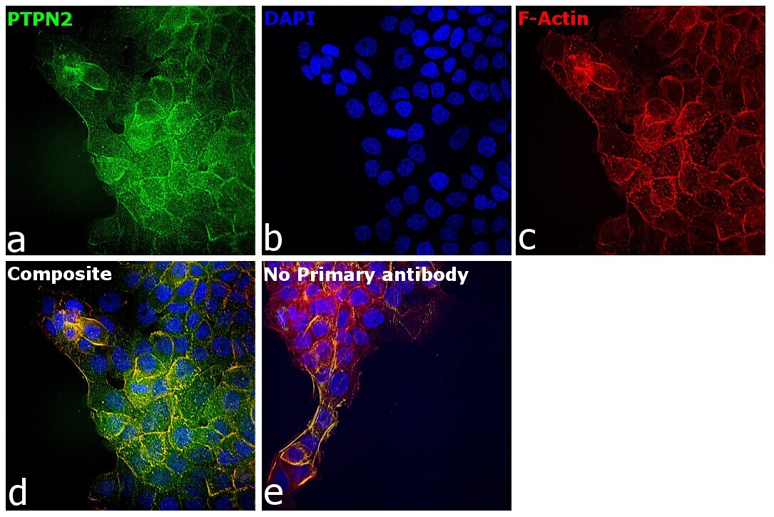
- Immunofluorescence analysis of PTPN2 was performed using 70% confluent log phase U-87 MG cells. The cells were fixed with 4% paraformaldehyde for 10 minutes, permeabilized with 0.01% Triton™ X-100 for 15 minutes, and blocked with 2% BSA for 1 hour at room temperature. The cells were labeled with PTPN2 Polyclonal Antibody (Product # PA5-42722) at 1:100 dilution in 0.1% BSA, incubated at 4 degree celsius overnight and then with Goat anti-Rabbit IgG (H+L) Highly Cross-Adsorbed Secondary Antibody, Alexa Fluor Plus 488 (Product # A27034) at a dilution of 1:2000 dilution for 45 minutes at room temperature (Panel a: green). Nuclei (Panel b: blue) were stained with Hoechst 33342 (Product # H1399). F-actin (Panel c: red) was stained with Rhodamine Phalloidin (Product # R415, 1:300 dilution). Panel d represents the merged image showing membrane-like localization for PTPN2 in U-87 MG. Panel e represents control cells with no primary antibody to assess background. The images were captured at 40X magnification in CellInsight CX7 LZR High-Content Screening (HCS) Platform (Product # CX7C1115LZR).

Supportive validation
- Submitted by
- Invitrogen Antibodies (provider)
- Main image
- Experimental details
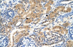
- Immunohistochemistry analysis of human kidney tissue using an anti-PTPN2 / TCPTP polyclonal antibody (Product # PA5-42722).
