Explore
 Validate
 Learn
Learn Western blot
Antibody data
- Antibody Data
- Antigen structure
- References [2]
- Comments [0]
- Validations
- Western blot [1]
- Immunocytochemistry [1]
- Other assay [1]
Submit
Validation data
Reference
Comment
Report error
- Product number
- 710535 - Provider product page

- Provider
- Invitrogen Antibodies
- Product name
- Phospho-Caspase 8 (Ser387) Recombinant Polyclonal Antibody (10HCLC)
- Antibody type
- Polyclonal
- Antigen
- Other
- Description
- Recombinant rabbit polyclonal antibodies are unique offerings from Thermo Fisher Scientific. They are comprised of a selection of multiple different recombinant monoclonal antibodies, providing the best of both worlds - the sensitivity of polyclonal antibodies with the specificity of monoclonal antibodies - all delivered with the consistency only found in a recombinant antibody. While functionally the same as a polyclonal antibody - recognizing multiple epitope sites on the target and producing higher detection sensitivity for low abundance targets - a recombinant rabbit polyclonal antibody has a known mixture of light and heavy chains. The exact population can be produced in every lot, circumventing the biological variability typically associated with polyclonal antibody production.
- Reactivity
- Human
- Host
- Rabbit
- Isotype
- IgG
- Antibody clone number
- 10HCLC
- Vial size
- 100 µg
- Concentration
- 0.5 mg/mL
- Storage
- Store at 4°C short term. For long term storage, store at -20°C, avoiding freeze/thaw cycles.
Submitted references Parthenolide inhibits the proliferation and induces the apoptosis of human uveal melanoma cells.
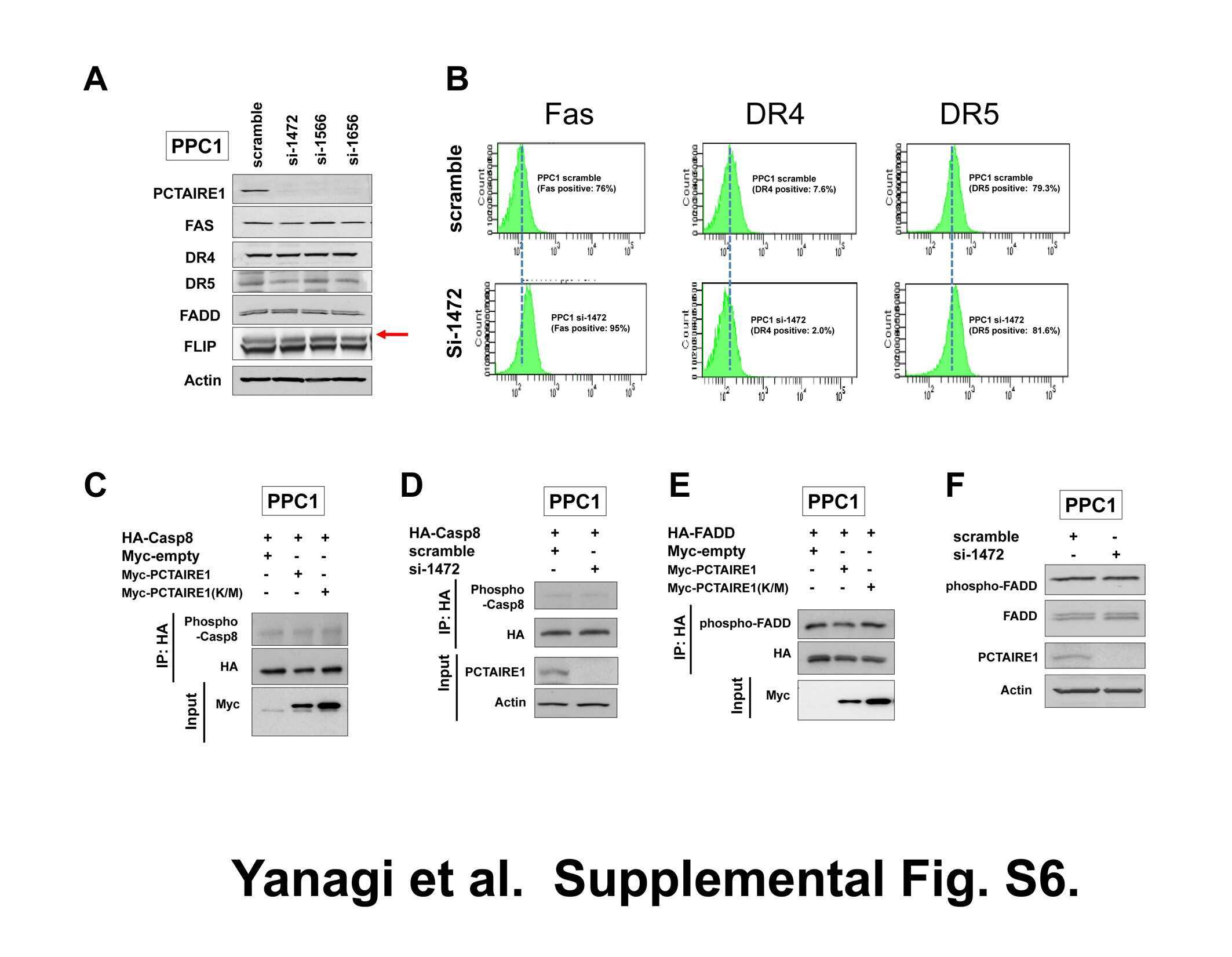
PCTAIRE1-knockdown sensitizes cancer cells to TNF family cytokines.
Che ST, Bie L, Li X, Qi H, Yu P, Zuo L
International journal of ophthalmology 2019;12(10):1531-1538
International journal of ophthalmology 2019;12(10):1531-1538
PCTAIRE1-knockdown sensitizes cancer cells to TNF family cytokines.
Yanagi T, Shi R, Aza-Blanc P, Reed JC, Matsuzawa S
PloS one 2015;10(3):e0119404
PloS one 2015;10(3):e0119404
No comments: Submit comment
Supportive validation
- Submitted by
- Invitrogen Antibodies (provider)
- Main image

- Experimental details
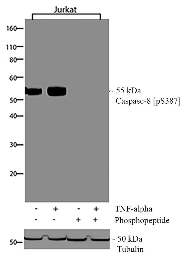
- Western blot analysis of Caspase-8 (pS387) was performed by loading 20 µg of Jurkat and Jurkat treated with TNF-alpha (20 ng/mL for 30 minutes) lysates (lane 1, 2) using Novex®NuPAGE®4-12% Bis-Tris gel (Product # NP0321BOX), XCell SureLock Electrophoresis System (Product # EI0002), Novex® Sharp Pre-Stained Protein Standard (Product # LC5800), and iBlot® Dry Blotting System (Product # IB21001). Proteins were transferred to a nitrocellulose membrane and blocked with 5% skim milk for 1 hour at room temperature. Caspase-8 (pS387) was detected at ~55 kDa using Caspase-8 (pS387) Recombinant Rabbit Polyclonal Antibody (Product # 710535) at 1:1000 dilution in 2.5% skim milk at 4°C overnight on a rocking platform. To confirm specificity, competition was performed with the phosphopeptide (10 µg/mL) (lane 3, 4). Goat anti-Rabbit IgG - HRP Secondary Antibody (Product # G-21234) at 1:5000 dilution was used and chemiluminescent detection was performed using Pierce™ ECL Western blotting Substrate (Product # 32106).
Supportive validation
- Submitted by
- Invitrogen Antibodies (provider)
- Main image

- Experimental details
- Immunofluorescent analysis of Caspase-8 (pS387) was done by treating HeLa cells with TNF-alpha at 20 ng/mL for 20 minutes. The cells were fixed with 4% paraformaldehyde for 15 minutes, permeabilized with 0.25% Triton X-100 for 10 minutes, and blocked with 5% BSA for 1 hour at room temperature. The cells were labeled with Caspase-8 (pS387) Recombinant Rabbit Polyclonal Antibody (Product # 710535) at a dilution of 1:500 in 1% BSA and incubated for 3 hours at room temperature and then labeled with Alexa Fluor® 488 Goat anti-Rabbit IgG Secondary Antibody (Product # A-11008) at a dilution of 1:400 for 30 minutes at room temperature (Panel a: green). F-actin (Panel b: red) was stained with Alexa Fluor® 594 Phalloidin (Product # A12381) and nuclei (Panel c: blue) were stained with SlowFade® Gold Antifade Mountant with DAPI (Product # S36938). Panel d is a merged image showing nuclear localization respectively and e shows competition with the phospho- Caspase 8 (pS387) peptide. The images were captured using a Nikon microscope at 20X magnification.

Supportive validation
- Submitted by
- Invitrogen Antibodies (provider)
- Main image
- Experimental details
- NULL