Explore
 Validate
 Learn
Learn Flow cytometry
Antibody data
- Antibody Data
- Antigen structure
- References [3]
- Comments [0]
- Validations
- Flow cytometry [1]
- Other assay [2]
Submit
Validation data
Reference
Comment
Report error
- Product number
- 12-9006-41 - Provider product page

- Provider
- Invitrogen Antibodies
- Product name
- Phospho-ZAP70/Syk (Tyr319, Tyr352) Monoclonal Antibody (n3kobu5), PE, eBioscience™
- Antibody type
- Monoclonal
- Antigen
- Other
- Description
- Description: This n3kobu5 monoclonal antibody recognizes human and mouse zeta chain-associated protein of 70 kD (also known as ZAP-70) and spleen tyrosine kinase (also known as SYK) when phosphorylated on Y319 and Y352, respectively. ZAP-70 and SYK are members of the SYK protein tyrosine kinase (PTK) family that are phosphorylated and activated by Src family PTKs. ZAP-70/SYK Y319/Y352 are located in the so-called interdomain of ZAP-70/SYK (between the N-terminal dual SH2 domains and the C-terminal kinase domain).
- Conjugate
- Yellow dye
- Antibody clone number
- n3kobu5
- Concentration
- 5 µL/Test
Submitted references CD19-CAR-T Cells Bearing a KIR/PD-1-Based Inhibitory CAR Eradicate CD19(+)HLA-C1(-) Malignant B Cells While Sparing CD19(+)HLA-C1(+) Healthy B Cells.
Long-Term Programming of CD8 T Cell Immunity by Perinatal Exposure to Glucocorticoids.
Neurokinin-1 Receptor Signaling Is Required for Efficient Ca(2+) Flux in T-Cell-Receptor-Activated T Cells.
Tao L, Farooq MA, Gao Y, Zhang L, Niu C, Ajmal I, Zhou Y, He C, Zhao G, Yao J, Liu M, Jiang W
Cancers 2020 Sep 13;12(9)
Cancers 2020 Sep 13;12(9)
Long-Term Programming of CD8 T Cell Immunity by Perinatal Exposure to Glucocorticoids.
Hong JY, Lim J, Carvalho F, Cho JY, Vaidyanathan B, Yu S, Annicelli C, Ip WKE, Medzhitov R
Cell 2020 Mar 5;180(5):847-861.e15
Cell 2020 Mar 5;180(5):847-861.e15
Neurokinin-1 Receptor Signaling Is Required for Efficient Ca(2+) Flux in T-Cell-Receptor-Activated T Cells.
Morelli AE, Sumpter TL, Rojas-Canales DM, Bandyopadhyay M, Chen Z, Tkacheva O, Shufesky WJ, Wallace CT, Watkins SC, Berger A, Paige CJ, Falo LD Jr, Larregina AT
Cell reports 2020 Mar 10;30(10):3448-3465.e8
Cell reports 2020 Mar 10;30(10):3448-3465.e8
No comments: Submit comment
Supportive validation
- Submitted by
- Invitrogen Antibodies (provider)
- Main image

- Experimental details
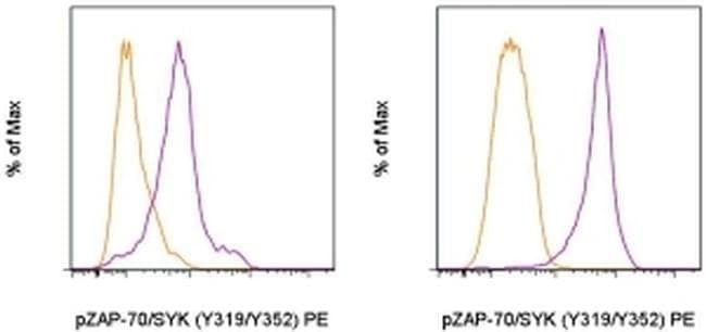
- Intracellular staining of untreated (orange histogram) or 5-minute hydrogen peroxide-activated sodium pervanadate-treated (purple histogram) mouse spleen cells with Anti-Human/Mouse phospho-ZAP-70/SYK (Y319/Y352) PE using the Intracellular Fixation & Permeabilization Buffer Set (Product # 88-8824-00) and protocol. Lymphocytes in the CD3+ (left) or B220+ (right) gates were used for analysis.
- Conjugate
- Yellow dye
Supportive validation
- Submitted by
- Invitrogen Antibodies (provider)
- Main image

- Experimental details
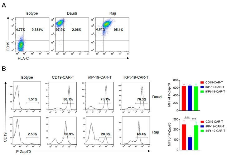
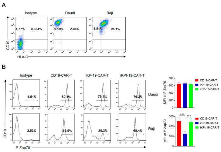
- Figure 2 Dephosphorylating P-Zap70 by iKP CAR via intracellular PD-1 domain. ( A ) Flow cytometric analysis of CD19 and HLA-C1 expression in Daudi cells or Raji cells by using APC-anti-human CD19 and PE-anti-human HLA-C antibodies. ( B ) Expression analysis of P-Zap70 in different CAR-T cells by flow cytometry. iKP-19-CAR-T/iKPt-19-CAR-T cells and CD19-CAR-T cells were exposed to Daudi cells or Raji cells for 6 h at a 1:1 ratio in RPMI-1640 medium, stained with PE-anti-human P-Zap70 antibody and MFI of P-Zap70 was statistically analyzed ( n = 4 different donors). All the experiments were conducted in triplicate manner using PBMCs from each donor. *** p < 0.001. Error bars represent +- SD. The CD19 CAR positive rate was unified using UT cells in all the co-culture experiments in this study.
- Conjugate
- Yellow dye
- Submitted by
- Invitrogen Antibodies (provider)
- Main image
- Experimental details
- Figure 2 Dephosphorylating P-Zap70 by iKP CAR via intracellular PD-1 domain. ( A ) Flow cytometric analysis of CD19 and HLA-C1 expression in Daudi cells or Raji cells by using APC-anti-human CD19 and PE-anti-human HLA-C antibodies. ( B ) Expression analysis of P-Zap70 in different CAR-T cells by flow cytometry. iKP-19-CAR-T/iKPt-19-CAR-T cells and CD19-CAR-T cells were exposed to Daudi cells or Raji cells for 6 h at a 1:1 ratio in RPMI-1640 medium, stained with PE-anti-human P-Zap70 antibody and MFI of P-Zap70 was statistically analyzed ( n = 4 different donors). All the experiments were conducted in triplicate manner using PBMCs from each donor. *** p < 0.001. Error bars represent +- SD. The CD19 CAR positive rate was unified using UT cells in all the co-culture experiments in this study.
- Conjugate
- Yellow dye