Explore
 Validate
 Learn
Learn Flow cytometry
 Other assay
Antibody data
- Antibody Data
- Antigen structure
- References [8]
- Comments [0]
- Validations
- Other assay [3]
Submit
Validation data
Reference
Comment
Report error
- Product number
- MHCD4501-4 - Provider product page

- Provider
- Invitrogen Antibodies
- Product name
- CD45 Monoclonal Antibody (HI30), FITC
- Antibody type
- Monoclonal
- Antigen
- Other
- Description
- Commonly used, FITC conjugates provide relatively high absorptivity, excellent fluorescence quantum yield, and good water solubility.
- Reactivity
- Human
- Host
- Mouse
- Conjugate
- Green dye
- Isotype
- IgG
- Antibody clone number
- HI30
- Vial size
- 2 mL
- Storage
- 4° C, store in dark
Submitted references Autologous Fractionated Adipose Tissue as a Natural Biomaterial and Novel One-Step Stem Cell Therapy for Repairing Articular Cartilage Defects.
Comparison of Volumetric and Bead-Based Counting of CD34 Cells by Single-Platform Flow Cytometry.
Phenotype and multipotency of rabbit (Oryctolagus cuniculus) amniotic stem cells.
Dynamic change in natural killer cell type in the human ocular mucosa in situ as means of immune evasion by adenovirus infection.
Dielectrophoretic capture and genetic analysis of single neuroblastoma tumor cells.
Unique phenotype of human uterine NK cells and their regulation by endogenous TGF-beta.
Evaluation of leukocyte stabilisation in TransFix-treated blood samples by flow cytometry and transmission electron microscopy.
Cell-derived microparticles circulate in healthy humans and support low grade thrombin generation.
Li Q, Zhao F, Li Z, Duan X, Cheng J, Zhang J, Fu X, Zhang J, Shao Z, Guo Q, Hu X, Ao Y
Frontiers in cell and developmental biology 2020;8:694
Frontiers in cell and developmental biology 2020;8:694
Comparison of Volumetric and Bead-Based Counting of CD34 Cells by Single-Platform Flow Cytometry.
Saraiva L, Wang L, Kammel M, Kummrow A, Atkinson E, Lee JY, Yalcinkaya B, Akgöz M, Höckner J, Ruf A, Engel A, Zhang YZ, O'Shea O, Sassi MP, Divieto C, Lekishvili T, Campbell J, Liu Y, Wang J, Stebbings R, Gaigalas AK, Rigsby P, Neukammer J, Vessillier S
Cytometry. Part B, Clinical cytometry 2019 Nov;96(6):508-513
Cytometry. Part B, Clinical cytometry 2019 Nov;96(6):508-513
Phenotype and multipotency of rabbit (Oryctolagus cuniculus) amniotic stem cells.
Borghesi J, Mario LC, Carreira AC, Miglino MA, Favaron PO
Stem cell research & therapy 2017 Feb 7;8(1):27
Stem cell research & therapy 2017 Feb 7;8(1):27
Dynamic change in natural killer cell type in the human ocular mucosa in situ as means of immune evasion by adenovirus infection.
Yawata N, Selva KJ, Liu YC, Tan KP, Lee AW, Siak J, Lan W, Vania M, Arundhati A, Tong L, Li J, Mehta JS, Yawata M
Mucosal immunology 2016 Jan;9(1):159-70
Mucosal immunology 2016 Jan;9(1):159-70
Dielectrophoretic capture and genetic analysis of single neuroblastoma tumor cells.
Carpenter EL, Rader J, Ruden J, Rappaport EF, Hunter KN, Hallberg PL, Krytska K, O'Dwyer PJ, Mosse YP
Frontiers in oncology 2014;4:201
Frontiers in oncology 2014;4:201
Unique phenotype of human uterine NK cells and their regulation by endogenous TGF-beta.
Eriksson M, Meadows SK, Wira CR, Sentman CL
Journal of leukocyte biology 2004 Sep;76(3):667-75
Journal of leukocyte biology 2004 Sep;76(3):667-75
Evaluation of leukocyte stabilisation in TransFix-treated blood samples by flow cytometry and transmission electron microscopy.
Canonico B, Zamai L, Burattini S, Granger V, Mannello F, Gobbi P, Felici C, Falcieri E, Reilly JT, Barnett D, Papa S
Journal of immunological methods 2004 Dec;295(1-2):67-78
Journal of immunological methods 2004 Dec;295(1-2):67-78
Cell-derived microparticles circulate in healthy humans and support low grade thrombin generation.
Berckmans RJ, Nieuwland R, Böing AN, Romijn FP, Hack CE, Sturk A
Thrombosis and haemostasis 2001 Apr;85(4):639-46
Thrombosis and haemostasis 2001 Apr;85(4):639-46
No comments: Submit comment
Supportive validation
- Submitted by
- Invitrogen Antibodies (provider)
- Main image
- Experimental details
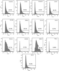
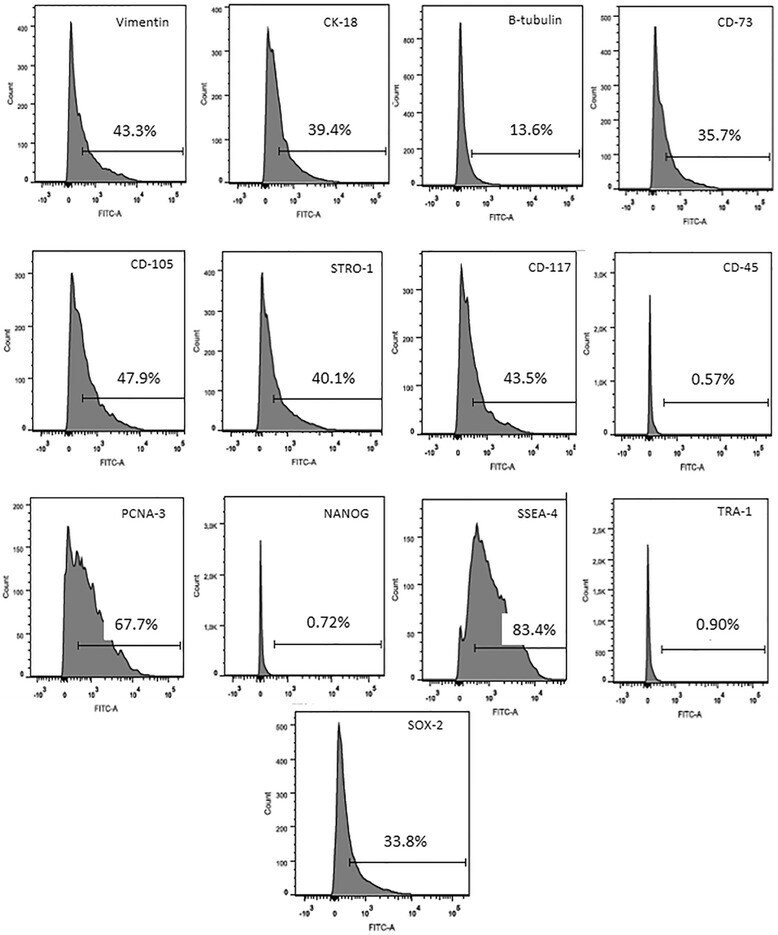
- Fig. 3 Immunophenotyping of rabbit amniotic cells at passage 8 analysed by flow cytometry. Note the expression levels of cytoskeletal markers (vimentin, cytokeratin and beta-tubulin) and mesenchymal cell markers (CD73, CD105 and Stro-1). CD117 (a marker of haematopoietic stem cell precursors) was highly expressed, while CD45 (a marker of haematopoietic cells) was not expressed. There were also significant levels of expression for the proliferation marker PCNA-3 and the pluripotency markers SSEA-4 and Sox-2, while other pluripotency markers (Nanog and TRA-1) were not expressed. FITC fluorescein isothiocyanate
- Conjugate
- Green dye
- Submitted by
- Invitrogen Antibodies (provider)
- Main image
- Experimental details
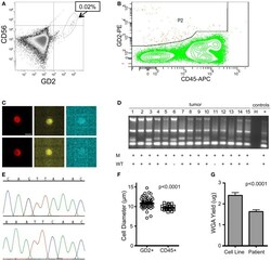
- Figure 5 Isolation and targeted sequencing of rare individual patient DTC . (A) The post-Ficoll fraction for a bone marrow sample from patient CHOP7 was fixed, stained for CD45, CD56, and GD2, and an aliquot was analyzed by flow cytometry. Given that only 0.02% of cells were determined to be GD2-positive, (B) pre-enrichment was conducted on the FACSAria using the wide P2 gate shown. (C) Representative images of staining for GD2 (first column; bar depicts 10 mum), CD56 (middle column), and CD45 (right column) are shown for single cells #15 (top row) and #7 (bottom row). (D) A gel for the quality control panel of four housekeeping genes is shown with a ""+"" below depicting detection of either the mutant allele (top row labeled ""M"" or the wild-type allele bottom row labeled ""WT""). (E) The chromatogram of the wild-type (top) and mutant allele (bottom) for cell #8. (F) Diameter of patient cells, as measured for 56 tumor cells and 22 WBCs while in solution on the DEPArray chip. (G) Bone marrow samples of patients were processed as described above and direct fluorescent staining used to measure the DNA concentration of WGA product from single cells. Mean WGA yield for n = 64 patient single cells is shown in comparison to the yield for n = 31 single tumor cells from cell line spiking experiments. In this analysis, all patient and cell line cells were fixed.
- Conjugate
- Green dye

- Submitted by
- Invitrogen Antibodies (provider)
- Main image
- Experimental details
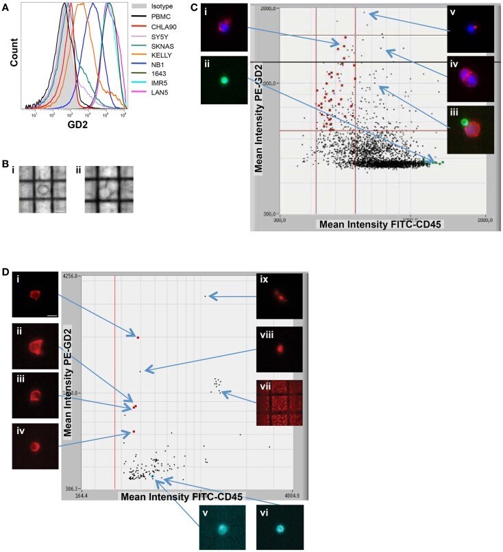
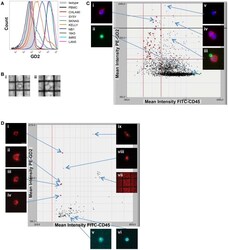
- Figure 2 Image-based capture of individual spiked cells . (A) Cell surface GD2 expression was measured by flow cytometry for the eight neuroblastoma cell lines used for isolation of single cells (and listed at right), as compared to negative control WBCs and isotype control. (B) WBCs and the GD2-dim neuroblastoma cell line SY5Y were mixed at a tumor:WBC ratio of 1:35, pre-labeled with GD2-PE, CD45-FITC, and Hoechst nuclear dye, then injected into the DEPArray. Shown are bright-field images of (i) a single tumor cell (bar depicts 10 mum) and (ii) a cluster of two cells. In (C) , a scatter plot of GD2-PE and CD45-FITC mean fluorescence intensity (MFI) is shown as well as cell images, including on the left-hand side of the dot-plot (i) an image of a single tumor cell and (ii) a single WBC, and on the right-hand side (iii) a heterogeneous cluster, (iv) a homogeneous cluster, and (v) a spurious event. (D) NB1643M cells and normal donor WBCs were mixed at a ratio of 1:1,000,000 and stained with GD2-PE and CD45-FITC, in this representative experiment. The sample was pre-enriched using FACSAria-based sorting, and the enriched fraction placed into the DEPArray cartridge. Shown are a scatter plot of GD2-PE and CD45-FITC MFI, as well as images of (i-iv) intact tumor cells, (v and vi) WBCs, and (vii-ix) debris and false-positive events.
- Conjugate
- Green dye