Explore
 Validate
 Learn
Learn Flow cytometry
Antibody data
- Antibody Data
- Antigen structure
- References [21]
- Comments [0]
- Validations
- Flow cytometry [1]
- Other assay [15]
Submit
Validation data
Reference
Comment
Report error
- Product number
- 56-9459-41 - Provider product page

- Provider
- Invitrogen Antibodies
- Product name
- CD45 Monoclonal Antibody (2D1), Alexa Fluor™ 700, eBioscience™
- Antibody type
- Monoclonal
- Antigen
- Other
- Description
- Description: The 2D1 monoclonal antibody reacts with all isoforms of human CD45, also known as Leukocyte Common Antigen (LCA). CD45 is expressed by all hematopoietic cells excluding circulating erythrocytes and platelets. The cytoplasmic portion of CD45 has tyrosine phosphatase enzymatic activity and plays an important role in activation of lymphocytes.
- Conjugate
- Near infrared dye
- Antibody clone number
- 2D1
- Concentration
- 5 µL/Test
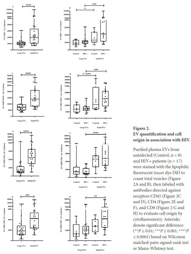
Submitted references Plasma Extracellular Vesicle Subtypes May be Useful as Potential Biomarkers of Immune Activation in People With HIV.
Bcl-xL mutant promotes cartilage differentiation of BMSCs by upregulating TGF-β/BMP expression levels.
Smac-mimetics reduce numbers and viability of human osteoclasts.
Heterogeneous disease-propagating stem cells in juvenile myelomonocytic leukemia.
VAP-PLGA microspheres (VAP-PLGA) promote adipose-derived stem cells (ADSCs)-induced wound healing in chronic skin ulcers in mice via PI3K/Akt/HIF-1α pathway.
Down-Regulated Exosomal MicroRNA-221 - 3p Derived From Senescent Mesenchymal Stem Cells Impairs Heart Repair.
Biological Therapy in Inflammatory Bowel Disease Patients Partly Restores Intestinal Innate Lymphoid Cell Subtype Equilibrium.
Single residue in CD28-costimulated CAR-T cells limits long-term persistence and antitumor durability.
Sodium Selenite Improves The Therapeutic Effect Of BMSCs Via Promoting The Proliferation And Differentiation, Thereby Promoting The Hematopoietic Factors.
TGF‑β induces periodontal ligament stem cell senescence through increase of ROS production.
High-yield isolation of menstrual blood-derived endometrial stem cells by direct red blood cell lysis treatment.
High Numbers of Circulating CD57(+) NK Cells Associate with Resistance to HER2-Specific Therapeutic Antibodies in HER2(+) Primary Breast Cancer.
Microglia innately develop within cerebral organoids.
A fully defined static suspension culture system for large-scale human embryonic stem cell production.
Similarities and differences between helminth parasites and cancer cell lines in shaping human monocytes: Insights into parallel mechanisms of immune evasion.
Increased expression of triggering receptor expressed on myeloid cells-1 in the population with obesity and insulin resistance.
Tumor immune microenvironment characterization in clear cell renal cell carcinoma identifies prognostic and immunotherapeutically relevant messenger RNA signatures.
Vascular niche promotes hematopoietic multipotent progenitor formation from pluripotent stem cells.
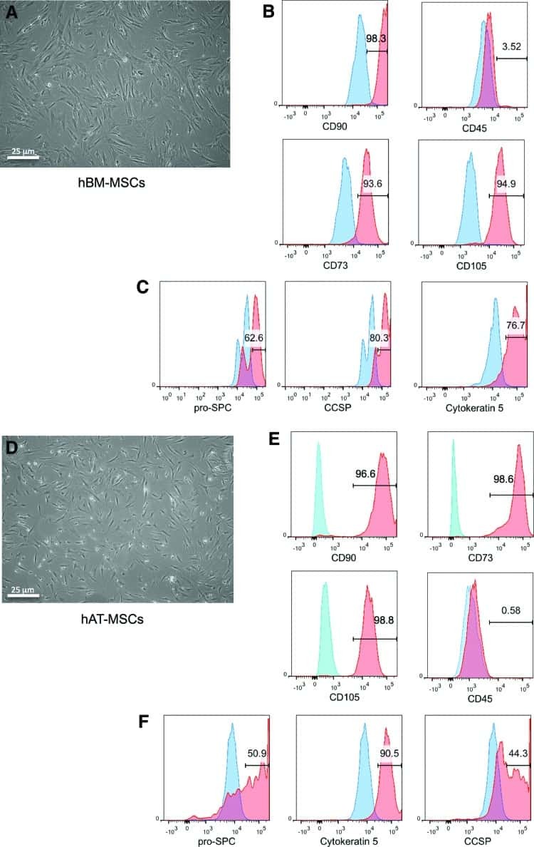
Epithelial cell differentiation of human mesenchymal stromal cells in decellularized lung scaffolds.
A deletion in the gene encoding the CD45 antigen in a patient with SCID.
Subpopulations of normal and leukemic human thymocytes: an analysis with the use of monoclonal antibodies.
Bazié WW, Boucher J, Vitry J, Goyer B, Routy JP, Tremblay C, Trottier S, Jenabian MA, Provost P, Alary M, Gilbert C
Pathogens & immunity 2021;6(1):1-28
Pathogens & immunity 2021;6(1):1-28
Bcl-xL mutant promotes cartilage differentiation of BMSCs by upregulating TGF-β/BMP expression levels.
Xiao K, Yang L, Xie W, Gao X, Huang R, Xie M
Experimental and therapeutic medicine 2021 Jul;22(1):736
Experimental and therapeutic medicine 2021 Jul;22(1):736
Smac-mimetics reduce numbers and viability of human osteoclasts.
Moen IN, Westhrin M, Håland E, Haug M, Nonstad U, Klaharn M, Standal T, Starheim KK
Cell death discovery 2021 Feb 19;7(1):36
Cell death discovery 2021 Feb 19;7(1):36
Heterogeneous disease-propagating stem cells in juvenile myelomonocytic leukemia.
Louka E, Povinelli B, Rodriguez-Meira A, Buck G, Wen WX, Wang G, Sousos N, Ashley N, Hamblin A, Booth CAG, Roy A, Elliott N, Iskander D, de la Fuente J, Fordham N, O'Byrne S, Inglott S, Norfo R, Salio M, Thongjuea S, Rao A, Roberts I, Mead AJ
The Journal of experimental medicine 2021 Feb 1;218(2)
The Journal of experimental medicine 2021 Feb 1;218(2)
VAP-PLGA microspheres (VAP-PLGA) promote adipose-derived stem cells (ADSCs)-induced wound healing in chronic skin ulcers in mice via PI3K/Akt/HIF-1α pathway.
Jiang W, Zhang J, Zhang X, Fan C, Huang J
Bioengineered 2021 Dec;12(2):10264-10284
Bioengineered 2021 Dec;12(2):10264-10284
Down-Regulated Exosomal MicroRNA-221 - 3p Derived From Senescent Mesenchymal Stem Cells Impairs Heart Repair.
Sun L, Zhu W, Zhao P, Zhang J, Lu Y, Zhu Y, Zhao W, Liu Y, Chen Q, Zhang F
Frontiers in cell and developmental biology 2020;8:263
Frontiers in cell and developmental biology 2020;8:263
Biological Therapy in Inflammatory Bowel Disease Patients Partly Restores Intestinal Innate Lymphoid Cell Subtype Equilibrium.
Creyns B, Jacobs I, Verstockt B, Cremer J, Ballet V, Vandecasteele R, Vanuytsel T, Ferrante M, Vermeire S, Van Assche G, Ceuppens JL, Breynaert C
Frontiers in immunology 2020;11:1847
Frontiers in immunology 2020;11:1847
Single residue in CD28-costimulated CAR-T cells limits long-term persistence and antitumor durability.
Guedan S, Madar A, Casado-Medrano V, Shaw C, Wing A, Liu F, Young RM, June CH, Posey AD Jr
The Journal of clinical investigation 2020 Jun 1;130(6):3087-3097
The Journal of clinical investigation 2020 Jun 1;130(6):3087-3097
Sodium Selenite Improves The Therapeutic Effect Of BMSCs Via Promoting The Proliferation And Differentiation, Thereby Promoting The Hematopoietic Factors.
Yan D, Tang B, Yan L, Zhang L, Miao M, Chen X, Sui G, Zhang Q, Liu D, Wang H
OncoTargets and therapy 2019;12:9685-9696
OncoTargets and therapy 2019;12:9685-9696
TGF‑β induces periodontal ligament stem cell senescence through increase of ROS production.
Fan C, Ji Q, Zhang C, Xu S, Sun H, Li Z
Molecular medicine reports 2019 Oct;20(4):3123-3130
Molecular medicine reports 2019 Oct;20(4):3123-3130
High-yield isolation of menstrual blood-derived endometrial stem cells by direct red blood cell lysis treatment.
Sun Y, Ren Y, Yang F, He Y, Liang S, Guan L, Cheng F, Liu Y, Lin J
Biology open 2019 May 2;8(5)
Biology open 2019 May 2;8(5)
High Numbers of Circulating CD57(+) NK Cells Associate with Resistance to HER2-Specific Therapeutic Antibodies in HER2(+) Primary Breast Cancer.
Muntasell A, Servitja S, Cabo M, Bermejo B, Pérez-Buira S, Rojo F, Costa-García M, Arpí O, Moraru M, Serrano L, Tusquets I, Martínez MT, Heredia G, Vera A, Martínez-García M, Soria L, Comerma L, Santana-Hernández S, Eroles P, Rovira A, Vilches C, Lluch A, Albanell J, López-Botet M
Cancer immunology research 2019 Aug;7(8):1280-1292
Cancer immunology research 2019 Aug;7(8):1280-1292
Microglia innately develop within cerebral organoids.
Ormel PR, Vieira de Sá R, van Bodegraven EJ, Karst H, Harschnitz O, Sneeboer MAM, Johansen LE, van Dijk RE, Scheefhals N, Berdenis van Berlekom A, Ribes Martínez E, Kling S, MacGillavry HD, van den Berg LH, Kahn RS, Hol EM, de Witte LD, Pasterkamp RJ
Nature communications 2018 Oct 9;9(1):4167
Nature communications 2018 Oct 9;9(1):4167
A fully defined static suspension culture system for large-scale human embryonic stem cell production.
Li X, Ma R, Gu Q, Liang L, Wang L, Zhang Y, Wang X, Liu X, Li Z, Fang J, Wu J, Wang Y, Li W, Hu B, Wang L, Zhou Q, Hao J
Cell death & disease 2018 Aug 30;9(9):892
Cell death & disease 2018 Aug 30;9(9):892
Similarities and differences between helminth parasites and cancer cell lines in shaping human monocytes: Insights into parallel mechanisms of immune evasion.
Narasimhan PB, Akabas L, Tariq S, Huda N, Bennuru S, Sabzevari H, Hofmeister R, Nutman TB, Tolouei Semnani R
PLoS neglected tropical diseases 2018 Apr;12(4):e0006404
PLoS neglected tropical diseases 2018 Apr;12(4):e0006404
Increased expression of triggering receptor expressed on myeloid cells-1 in the population with obesity and insulin resistance.
Subramanian S, Pallati PK, Rai V, Sharma P, Agrawal DK, Nandipati KC
Obesity (Silver Spring, Md.) 2017 Mar;25(3):527-538
Obesity (Silver Spring, Md.) 2017 Mar;25(3):527-538
Tumor immune microenvironment characterization in clear cell renal cell carcinoma identifies prognostic and immunotherapeutically relevant messenger RNA signatures.
Şenbabaoğlu Y, Gejman RS, Winer AG, Liu M, Van Allen EM, de Velasco G, Miao D, Ostrovnaya I, Drill E, Luna A, Weinhold N, Lee W, Manley BJ, Khalil DN, Kaffenberger SD, Chen Y, Danilova L, Voss MH, Coleman JA, Russo P, Reuter VE, Chan TA, Cheng EH, Scheinberg DA, Li MO, Choueiri TK, Hsieh JJ, Sander C, Hakimi AA
Genome biology 2016 Nov 17;17(1):231
Genome biology 2016 Nov 17;17(1):231
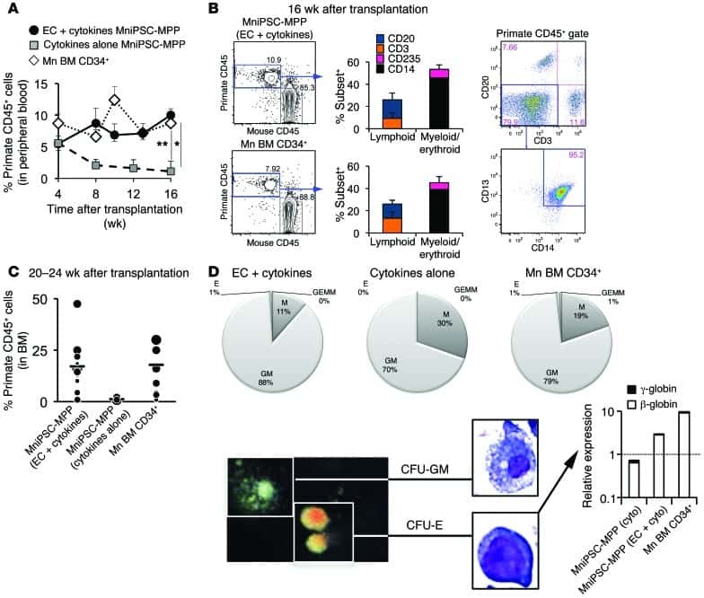
Vascular niche promotes hematopoietic multipotent progenitor formation from pluripotent stem cells.
Gori JL, Butler JM, Chan YY, Chandrasekaran D, Poulos MG, Ginsberg M, Nolan DJ, Elemento O, Wood BL, Adair JE, Rafii S, Kiem HP
The Journal of clinical investigation 2015 Mar 2;125(3):1243-54
The Journal of clinical investigation 2015 Mar 2;125(3):1243-54
Epithelial cell differentiation of human mesenchymal stromal cells in decellularized lung scaffolds.
Mendez JJ, Ghaedi M, Steinbacher D, Niklason LE
Tissue engineering. Part A 2014 Jun;20(11-12):1735-46
Tissue engineering. Part A 2014 Jun;20(11-12):1735-46
A deletion in the gene encoding the CD45 antigen in a patient with SCID.
Tchilian EZ, Wallace DL, Wells RS, Flower DR, Morgan G, Beverley PC
Journal of immunology (Baltimore, Md. : 1950) 2001 Jan 15;166(2):1308-13
Journal of immunology (Baltimore, Md. : 1950) 2001 Jan 15;166(2):1308-13
Subpopulations of normal and leukemic human thymocytes: an analysis with the use of monoclonal antibodies.
Bradstock KF, Janossy G, Pizzolo G, Hoffbrand AV, McMichael A, Pilch JR, Milstein C, Beverley P, Bollum FJ
Journal of the National Cancer Institute 1980 Jul;65(1):33-42
Journal of the National Cancer Institute 1980 Jul;65(1):33-42
No comments: Submit comment
Supportive validation
- Submitted by
- Invitrogen Antibodies (provider)
- Main image

- Experimental details
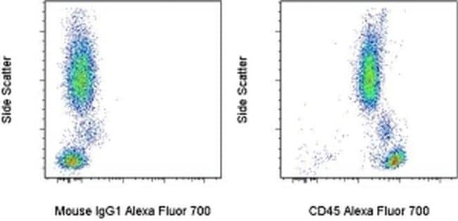
- Staining of normal human peripheral blood cells Mouse IgG1 K Isotype Control Alexa Fluor® 700 (Product # 56-4714-80) (left) or Anti-Human CD45 Alexa Fluor® 700 (right). Total viable cells were used for analysis.
- Conjugate
- Near infrared dye
Supportive validation
- Submitted by
- Invitrogen Antibodies (provider)
- Main image
- Experimental details
- NULL
- Conjugate
- Near infrared dye
- Submitted by
- Invitrogen Antibodies (provider)
- Main image

- Experimental details
- NULL
- Conjugate
- Near infrared dye
- Submitted by
- Invitrogen Antibodies (provider)
- Main image
- Experimental details
- NULL
- Conjugate
- Near infrared dye

- Submitted by
- Invitrogen Antibodies (provider)
- Main image

- Experimental details
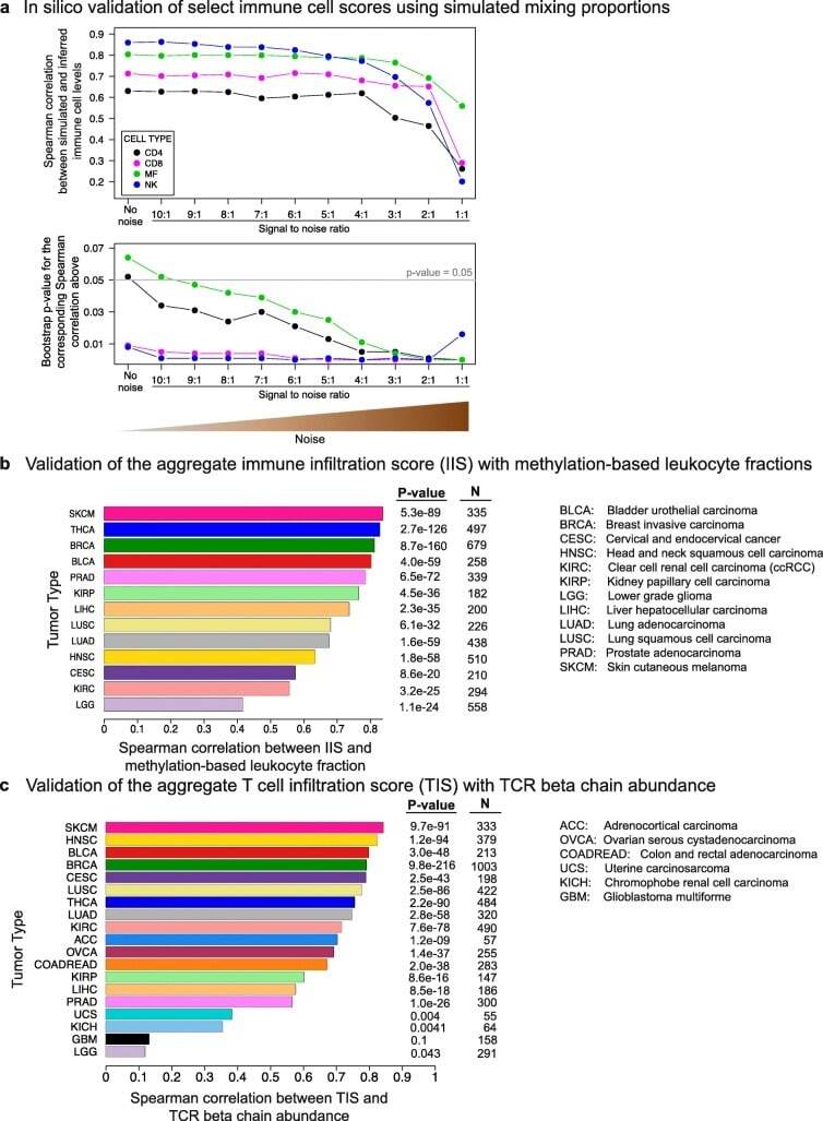
- Fig. 2 In silico validation of the immune cell scoring method. a In silico validation of immune cell scores using simulated mixing proportions. RNA-Seq profiles of FACS-sorted NK cells, macrophages, CD 4 + and CD 8 + T cells, and non-immune CD 45 - cells were mixed with known proportions to obtain a ""clean"" mixture. Noise was added at varying SNRs. Mixing levels were then inferred by ssGSEA from the ""clean"" and noisy mixtures. The Spearman correlations between the simulated and inferred levels ( top panel ) and the bootstrap p values for these correlation values ( bottom panel ) are shown on the y-axes (Additional file 1 : Figure S18 and "" Methods "" for the calculation of the bootstrap p values). b Validation of IIS with methylation-based leukocyte fractions. Spearman correlations between the two orthogonal scores are shown on the x-axis for 13 tumor types. c Validation of TIS with TCR beta chain abundance. Both scores are computationally inferred from RNA-Seq data but employ different approaches to measure T cell levels. Spearman correlations are shown on the x-axis for 19 tumor types
- Conjugate
- Near infrared dye
- Submitted by
- Invitrogen Antibodies (provider)
- Main image
- Experimental details
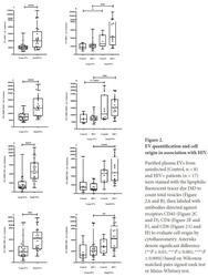
- Figure 2. EV quantification and cell origin in association with HIV. Purified plasma EVs fromuninfected (Control, n = 8) and HIV+ patients (n = 17) were stained with the lipophilic fluorescent tracer dye DiD to count total vesicles ( Figure 2A and B ), then labeled with antibodies directed against receptors CD45 ( Figure 2C and D ), CD4 ( Figure 2E and F ), and CD8 ( Figure 2 G and H ) to evaluate cell origin by cytofluorometry. Asterisks denote significant difference (** P
- Conjugate
- Near infrared dye
- Submitted by
- Invitrogen Antibodies (provider)
- Main image
- Experimental details
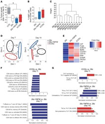
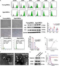
- FIGURE 1 Characterization of young and aged MSCs and exosomes. (A) Surface marker profiling of young-MSCs and aged-MSCs. (B) SA-beta-Gal staining showed that senescence increased significantly in aged MSCs. (C) Representative immunoblot images and quantitative analysis of p21, p53, and p16 protein level in young and aged-MSCs. ( n = 3). (D) Quantitation of cell cycle phases by propidium iodide staining. ( n = 3). (E) The CCK-8 assay showed that aged MSCs grew more slowly than young MSCs. ( n = 6). (F) Young and aged exosomes were observed using TEM. (G) The exosome surface markers were analyzed by Western blot. (H) Nanoparticle tracking analysis was used to analyze the particle size and concentration of Young-Exo and Aged-Exo. * p < 0.05; ** p < 0.01; *** p < 0.001; **** p < 0.0001; NS, not significant.
- Conjugate
- Near infrared dye
- Submitted by
- Invitrogen Antibodies (provider)
- Main image
- Experimental details
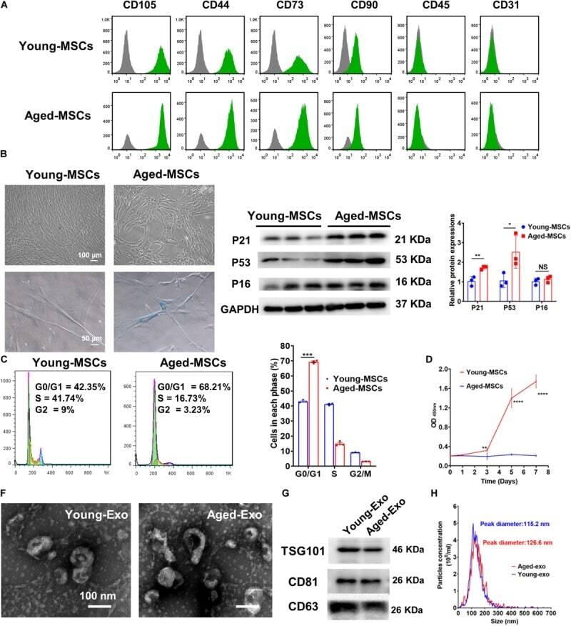
- Figure 1 Culture and identification of bone marrow mesenchymal stem cells. (A) Light microscopy of BMSCs (scale bar=100 um for the left image and 200 um for the right image). (B) Percentage of CD34-, CD45-, CD73-, CD90- and CD105-positive BMSCs were detected by flow cytometry. BMSC, bone marrow mesenchymal stem cell.
- Conjugate
- Near infrared dye

- Submitted by
- Invitrogen Antibodies (provider)
- Main image
- Experimental details
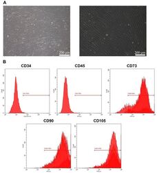
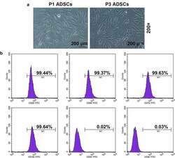
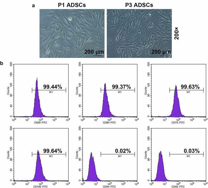
- Figure 1. Morphology and immune phenotype of adipose-derived stem cells (ADSCs) were identified by morphological observation and flow cytometry. (a) Morphology of the primary (P1) and third passage (P3) of ADSCs. Images were acquired at 200x magnification. (b) Immune phenotype of ADSCs. The average data from three independent experiments were shown as mean +- standard deviation
- Conjugate
- Near infrared dye
- Submitted by
- Invitrogen Antibodies (provider)
- Main image
- Experimental details
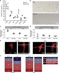
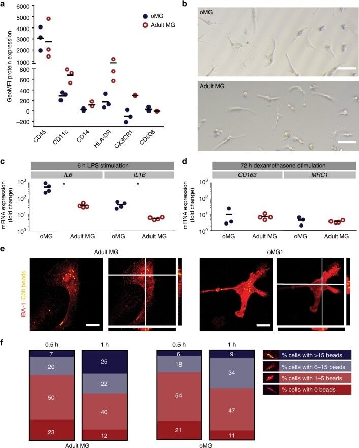
- Fig. 4 oMG expressed microglia-characteristic cell surface markers and showed similar functional immune and phagocytic properties as adult MG. a Flow cytometric analyses of the expression pattern of microglial extracellular markers on CD11b+-gated oMG (oMG 1, 3, and 5) compared to adult MG derived from three separate brain regions from adult MG1.1. (eight organoids were pooled per donor (oMG 1, 3, and 5) after 52 days in culture). b Morphology of magnetic automated cell sorted CD11b+ oMG 1 and adult MG in bright field microscope after 1 week in culture. Scale bar 40 mum. c mRNA expression, determined by qRT-PCR, of pro-inflammatory cytokines IL6 and IL1B after 6 h stimulation with LPS was significantly higher in oMG compared to adult MG (Mann-Whitney test IL6 and IL1B: U = 0, n = 4, p = 0.03). LPS-stimulated response relative to control condition without LPS. ( n = 4 experiments, eight organoids pooled per experiment; adult MG1.1) (* p < 0.05). d Anti-inflammatory response of oMG and adult MG was compared by qRT-PCR for expression of anti-inflammatory genes CD163 and MRC1 upon 72 h stimulation with dexamethasone. Dexamethasone-stimulated response relative to control condition without dexamethasone. (oMG, n = 3 separate experiments in which oMG were isolated from > 4 pooled cerebral organoids from iPSC 1 per experiment; adult MG, n = 4). e Phagocytosis capacity was tested oMG 1 and adult MG by performing a phagocytosis assay with iC3b-coated green-yellow
- Conjugate
- Near infrared dye
- Submitted by
- Invitrogen Antibodies (provider)
- Main image
- Experimental details
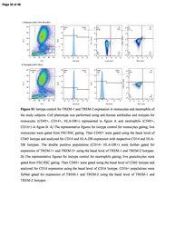
- 01 Figure S1 Isotype control for TREM-1 and TREM-2 expression in monocytes and neutrophils of the study subjects. Cell phenotype was performed using anti-human antibodies and isotypes for monocytes (CD45+, CD14+, HLA-DR+) represented in figure A and neutrophils (CD45+, CD16+) in figure B. A) The representative figures for isotype control for monocytes gating; live monocytes were gated from FSC/SSC gating. Then CD45+ were gated using the basal level of CD45 Isotype and analyzed for CD14 and HLA-DR expression with respective CD14 and HLA-DR Isotypes. The double positive populations (CD14+ HLA-DR+) were further gated for expression of TREM-1+ and TREM-2+ using the basal level of TREM-1 and TREM-2 Isotypes. B) The representative figures for Isotype control for neutrophils gating; live granulocytes were gated from FSC/SSC gating. Then CD45+ were gated using the basal level of CD45 Isotype and analyzed for CD16 expression using the basal level of CD16 Isotype. CD16+ populations were further gated for expression of TREM-1 and TREM-2 using the basal level of TREM-1 and TREM-2 Isotypes. Figure S2 Hematoxylin and Eosin staining for fatty liver grading and inflammation. H & E in biopsy samples of SO - D - , SO + D - and SO + D + groups respectively in liver (images Aa, Ab & Ac), omental fat (images Ba, Bb & Bc) and subcutaneous fat (images Ca, Cb & Cc). Liver biopsy samples showed steatosis in SO + D - and fibrosis and cirrhosis in SO + D + groups. Size of the adipocyte was larger in su
- Conjugate
- Near infrared dye
- Submitted by
- Invitrogen Antibodies (provider)
- Main image

- Experimental details
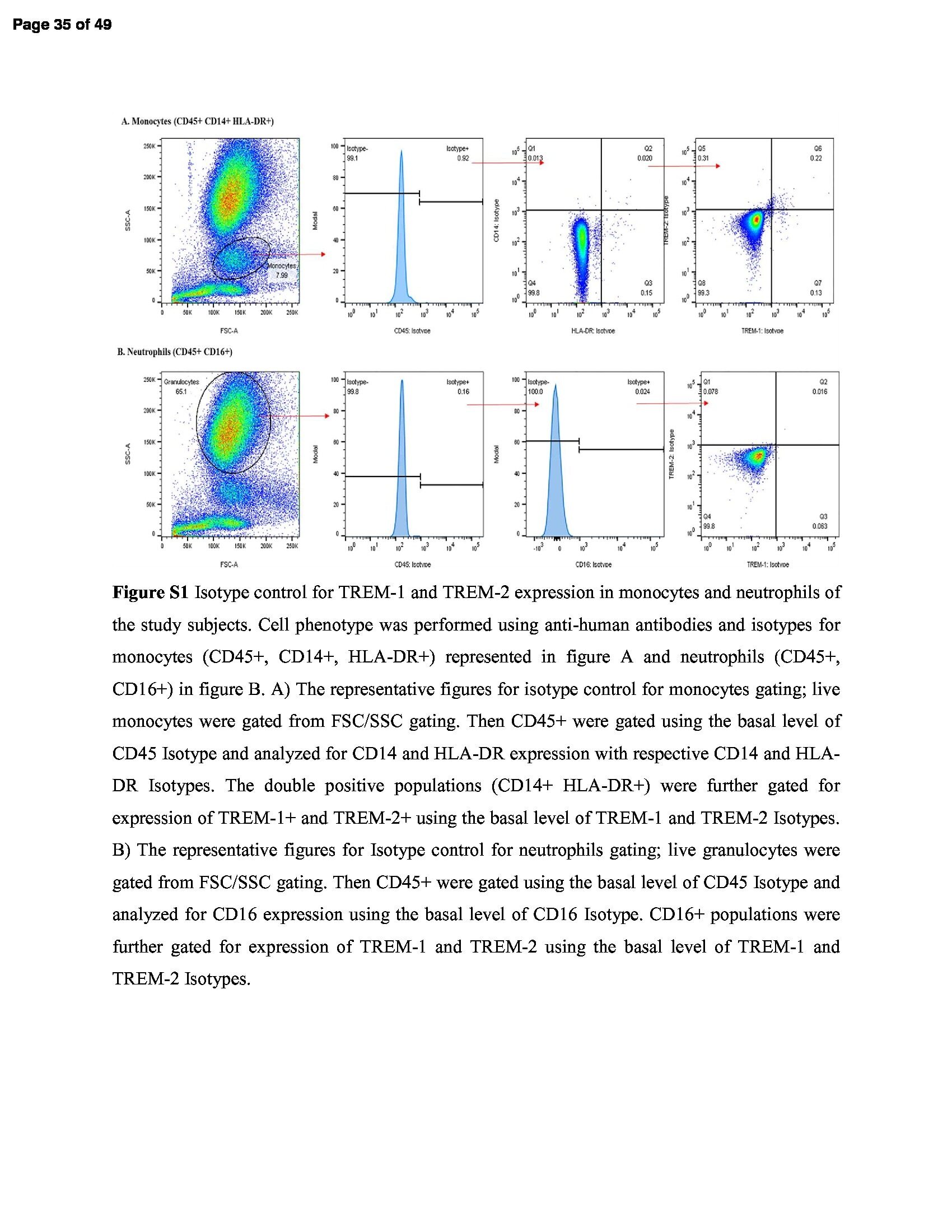
- Figure 1. Characterization of PDLSCs. (A) PDL cell clusters exhibited radiating or whirlpool-like morphology. The central structure in this image is a fragment of PDL tissue. Scale bar, 200 mum (B) CD146 + PDLSCs were small, round, fusiform and triangular. Scale bar, 100 mum. (C) PDLSCs were positive for the stem cell markers CD44, CD90 and CD105, but negative for CD34 and CD45, as detected by flow cytometry. PDL, periodontal ligament; PDLSCs, PDL stem cells.
- Conjugate
- Near infrared dye
- Submitted by
- Invitrogen Antibodies (provider)
- Main image
- Experimental details
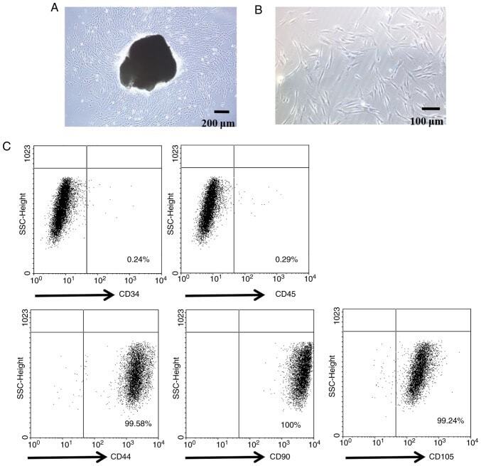
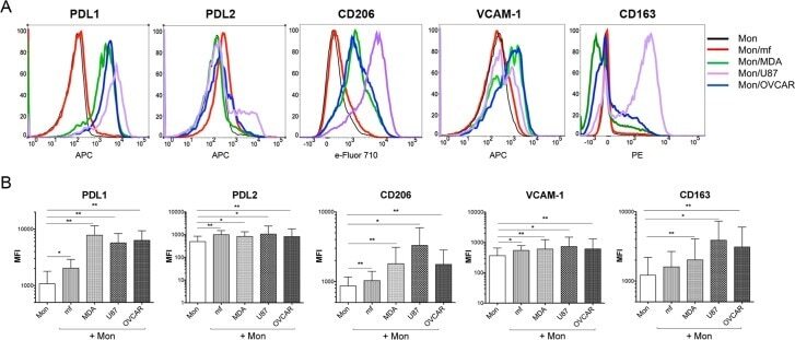
- Fig 3 Cancer cell lines and mf significantly upregulates the cell surface expressions of PDL1, PDL2, CD206 and VCAM-1 on human monocytes. Human monocytes were cultured in media alone, or with CMFDA-labeled three different cancer cell lines (MDA, OVCAR, U87), or with live mf of Brugia malayi for 48hr. Cells were harvested and cell surface expression PDL1, PDL2, CD206, VCAM-1, and CD163 was measured using flow cytometry gated on CD45 + /CMFDA - monocytes. (A) One representative set (n = 15) of flow histograms demonstrating cell surface expression in unexposed human monocytes and after exposure to mf or different cancer cell lines. (B). The data are expressed as the geometric mean with 95% confidence interval of the mean fluorescent intensity of unexposed and exposed monocytes ( n = 15). * P< 0.05, ** P< 0.005.
- Conjugate
- Near infrared dye
- Submitted by
- Invitrogen Antibodies (provider)
- Main image
- Experimental details
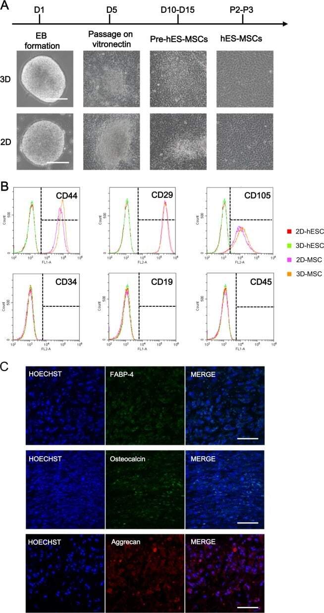
- Fig. 4 Comparison of MSCs derived from 2D-hESCs and 3D-hESCs. a The morphology of 2D- and 3D-hESC-MSCs at different stages of differentiation. b Flow cytometry analysis revealed specific MSC surface markers (CD44, CD29, and CD105) with negative controls (CD34, CD19, and CD45) in 2D- and 3D-hESC-MSCs. c Immunostaining of differentiated 3D-hESC-MSCs expressing an adipocyte marker (FABP-4), osteocytes maker (osteocalcin), and chondrocytes marker (aggrecan)
- Conjugate
- Near infrared dye
- Submitted by
- Invitrogen Antibodies (provider)
- Main image
- Experimental details
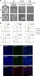
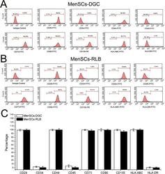
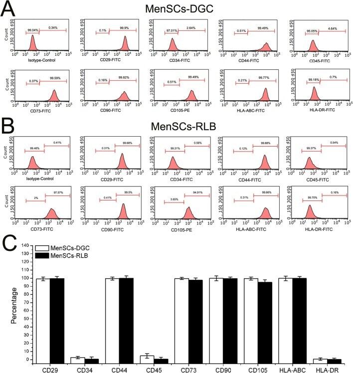
- Fig. 3. The phenotype of MenSCs. (A,B) To determine the immunophenotype of MenSCs, P3 MenSCs-DGC ( n =5) and MenSCs-RLB ( n =5) were stained by corresponding conjugated antibodies and analyzed by FACS. Both MenSCs-DGC and MenSCs-RLB positively expressed classical ASCs' markers (CD29, CD44, CD73, CD90 and CD105) and HLA-ABC; they did not express hematopoietic stem cell markers (CD34 and CD45) and HLA-DR. (C) The quantification of flow cytometry results of A and B.
- Conjugate
- Near infrared dye
- Submitted by
- Invitrogen Antibodies (provider)
- Main image
- Experimental details
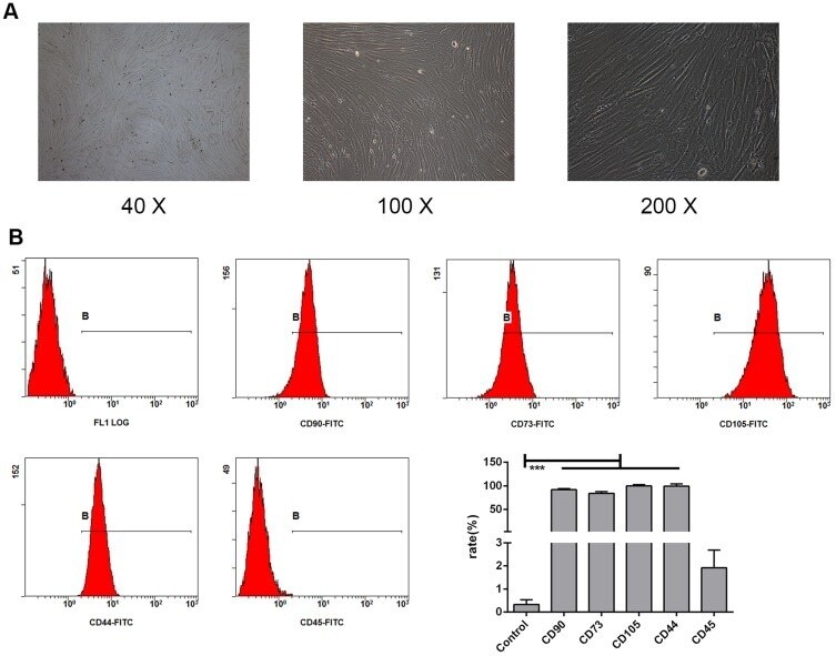
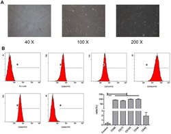
- Figure 1 Identification of BMSCs in mice. Morphological observations at different magnifications (40x, 100x, and 200x) showed that the bone marrow stem cells (BMSCs) isolated from mice were homogeneously elongated ( A ). Various cell surface markers of BMSCs were detected on the cells, including CD90, CD73, CD105, CD44, and CD45 ( B ). The values are from triplicate determinations. A P value less than 0.05 was considered to be statistically significant. ***Indicates a P value less than 0.001.
- Conjugate
- Near infrared dye