 Explore
 Validate
 Learn
Learn Immunohistochemistry
Antibody data
- Antibody Data
- Antigen structure
- References [9]
- Comments [0]
- Validations
- Immunohistochemistry [1]
- Other assay [7]
Submit
Validation data
Reference
Comment
Report error
- Product number
- 13-9457-82 - Provider product page

- Provider
- Invitrogen Antibodies
- Product name
- CD45 Monoclonal Antibody (CD45-2B11), Biotin, eBioscience™
- Antibody type
- Monoclonal
- Antigen
- Other
- Description
- Description: The CD45-2B11 monoclonal antibody reacts with human CD45, also known as Leukocyte Common Antigen (LCA). CD45 is expressed by all hematopoietic cells excluding circulating erythrocytes and platelets. The cytoplasmic portion of CD45 has tyrosine phosphatase enzymatic activity and plays an important role in lymphocyte proliferation and differentiation. The 2B11 antibody is useful for recognition of normal and neoplastic lymphoid cells.
- Conjugate
- Biotin
- Antibody clone number
- CD45-2B11
- Concentration
- 0.5 mg/mL
Submitted references Exosomes derived from stem cells of human deciduous exfoliated teeth inhibit angiogenesis in vivo and in vitro via the transfer of miR-100-5p and miR-1246.
Macrophage-derived IL-6 trans-signalling as a novel target in the pathogenesis of bronchopulmonary dysplasia.
Ex vivo culture of intact human patient derived pancreatic tumour tissue.
Heterogeneous nuclear ribonucleoprotein A2/B1 is a negative regulator of human breast cancer metastasis by maintaining the balance of multiple genes and pathways.
SHQ1 regulation of RNA splicing is required for T-lymphoblastic leukemia cell survival.
Adoptive transfer of xenoantigen‑stimulated T cell receptor Vβ‑restricted human regulatory T cells prevents porcine islet xenograft rejection in humanized mice.
Heterogeneous fibroblasts underlie age-dependent tertiary lymphoid tissues in the kidney.
Inflammatory malignant fibrous histiocytoma: distinction from Hodgkin's disease and non-Hodgkin's lymphoma by a panel of leukocyte markers.
Diagnosis of human lymphoma with monoclonal antileukocyte antibodies.
Liu P, Zhang Q, Mi J, Wang S, Xu Q, Zhuang D, Chen W, Liu C, Zhang L, Guo J, Wu X
Stem cell research & therapy 2022 Mar 3;13(1):89
Stem cell research & therapy 2022 Mar 3;13(1):89
Macrophage-derived IL-6 trans-signalling as a novel target in the pathogenesis of bronchopulmonary dysplasia.
Hirani D, Alvira CM, Danopoulos S, Milla C, Donato M, Tian L, Mohr J, Dinger K, Vohlen C, Selle J, V Koningsbruggen-Rietschel S, Barbarino V, Pallasch C, Rose-John S, Odenthal M, Pryhuber GS, Mansouri S, Savai R, Seeger W, Khatri P, Al Alam D, Dötsch J, Alejandre Alcazar MA
The European respiratory journal 2022 Feb;59(2)
The European respiratory journal 2022 Feb;59(2)
Ex vivo culture of intact human patient derived pancreatic tumour tissue.
Kokkinos J, Sharbeen G, Haghighi KS, Ignacio RMC, Kopecky C, Gonzales-Aloy E, Youkhana J, Timpson P, Pereira BA, Ritchie S, Pandzic E, Boyer C, Davis TP, Butler LM, Goldstein D, McCarroll JA, Phillips PA
Scientific reports 2021 Jan 21;11(1):1944
Scientific reports 2021 Jan 21;11(1):1944
Heterogeneous nuclear ribonucleoprotein A2/B1 is a negative regulator of human breast cancer metastasis by maintaining the balance of multiple genes and pathways.
Liu Y, Li H, Liu F, Gao LB, Han R, Chen C, Ding X, Li S, Lu K, Yang L, Tian HM, Chen BB, Li X, Xu DH, Deng XL, Shi SL
EBioMedicine 2020 Jan;51:102583
EBioMedicine 2020 Jan;51:102583
SHQ1 regulation of RNA splicing is required for T-lymphoblastic leukemia cell survival.
Su H, Hu J, Huang L, Yang Y, Thenoz M, Kuchmiy A, Hu Y, Li P, Feng H, Zhou Y, Taghon T, Van Vlierberghe P, Qing G, Chen Z, Liu H
Nature communications 2018 Oct 15;9(1):4281
Nature communications 2018 Oct 15;9(1):4281
Adoptive transfer of xenoantigen‑stimulated T cell receptor Vβ‑restricted human regulatory T cells prevents porcine islet xenograft rejection in humanized mice.
Jin X, Hu M, Gong L, Li H, Wang Y, Ji M, Li H
Molecular medicine reports 2018 Nov;18(5):4457-4467
Molecular medicine reports 2018 Nov;18(5):4457-4467
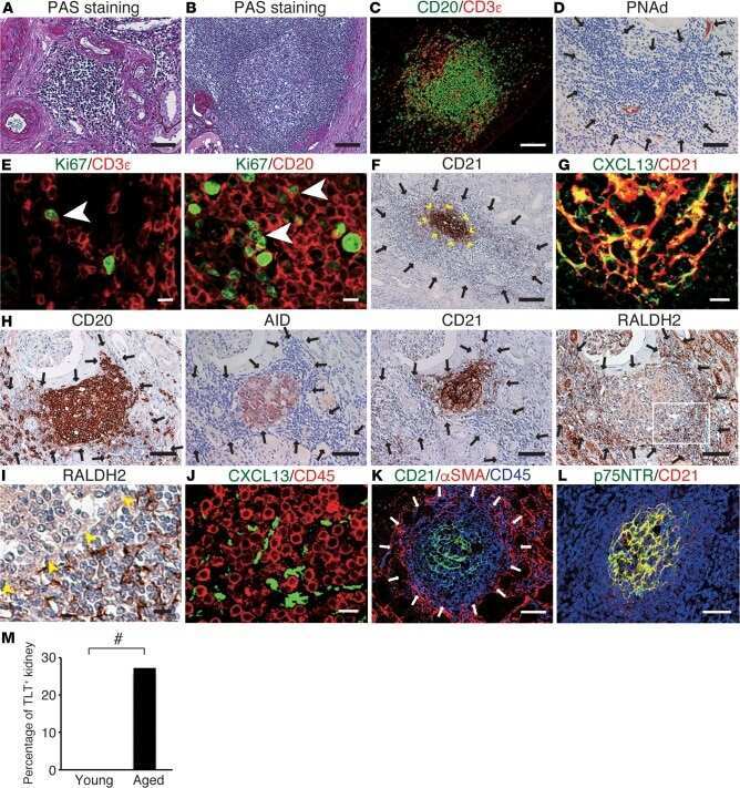
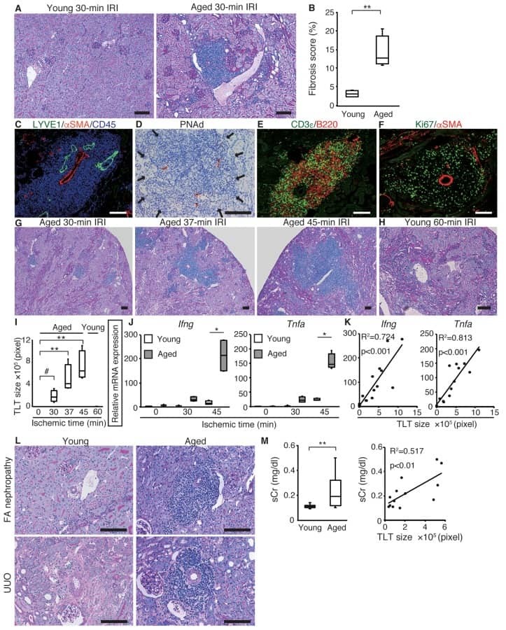
Heterogeneous fibroblasts underlie age-dependent tertiary lymphoid tissues in the kidney.
Sato Y, Mii A, Hamazaki Y, Fujita H, Nakata H, Masuda K, Nishiyama S, Shibuya S, Haga H, Ogawa O, Shimizu A, Narumiya S, Kaisho T, Arita M, Yanagisawa M, Miyasaka M, Sharma K, Minato N, Kawamoto H, Yanagita M
JCI insight 2016 Jul 21;1(11):e87680
JCI insight 2016 Jul 21;1(11):e87680
Inflammatory malignant fibrous histiocytoma: distinction from Hodgkin's disease and non-Hodgkin's lymphoma by a panel of leukocyte markers.
Khalidi HS, Singleton TP, Weiss SW
Modern pathology : an official journal of the United States and Canadian Academy of Pathology, Inc 1997 May;10(5):438-42
Modern pathology : an official journal of the United States and Canadian Academy of Pathology, Inc 1997 May;10(5):438-42
Diagnosis of human lymphoma with monoclonal antileukocyte antibodies.
Warnke RA, Gatter KC, Falini B, Hildreth P, Woolston RE, Pulford K, Cordell JL, Cohen B, De Wolf-Peeters C, Mason DY
The New England journal of medicine 1983 Nov 24;309(21):1275-81
The New England journal of medicine 1983 Nov 24;309(21):1275-81
No comments: Submit comment
Supportive validation
- Submitted by
- Invitrogen Antibodies (provider)
- Main image
- Experimental details
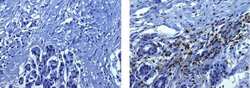
- Immunohistochemistry on formalin-fixed paraffin embedded human pancreas using 20 µg/mL of Mouse IgG1 K Isotype Control (left) or 20 µg/mL Anti-Human CD45 Biotin (right) followed by DAB visualization. Nuclei are counterstained with hematoxylin.
- Conjugate
- Biotin
Supportive validation
- Submitted by
- Invitrogen Antibodies (provider)
- Main image

- Experimental details
- NULL
- Conjugate
- Biotin
- Submitted by
- Invitrogen Antibodies (provider)
- Main image

- Experimental details
- NULL
- Conjugate
- Biotin

- Submitted by
- Invitrogen Antibodies (provider)
- Main image
- Experimental details
- NULL
- Conjugate
- Biotin
- Submitted by
- Invitrogen Antibodies (provider)
- Main image
- Experimental details
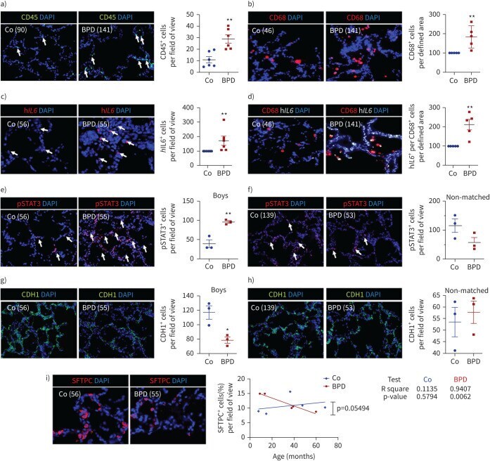
- FIGURE 9 Inflammation and interleukin 6 (IL-6)/signal transducer and activator of transcription 3 (STAT3) signalling in lungs of infants with bronchopulmonary dysplasia (BPD) and non-BPD. a) Representative immunofluorescent staining for immune cells using CD45 as a marker (green) in age-matched BPD and control lungs (Co); the lung identification numbers of the infants are indicated in brackets. Immune cells (CD45 + cells) were counted in 4-12 fields of view per lung. Summary data of the quantification of immune cells (CD45 + cells) per field of view for all infants (n=6 per group). DAPI: 4',6-diamidino-2-phenylindole. b) Representative co-immunofluorescent localisation of CD68 (marker of macrophages, red) in age-matched BPD and Co lungs; white arrows are indicating CD68 + cells. The analysis of CD68 + cells per 6-12 fields of view is shown next to the images; n=6 per group. c) Representative localisation of human IL6 (h IL6 , red) in lungs with BPD and Co; white arrows are indicating h IL6 + in situ hybridisation. The analysis of h IL6 + cells per 6-12 fields of view is shown next to the images; n=6 per group. d) Representative co-immunofluorescent localisation of CD68 (marker of macrophages, red) with h IL6 (white; in situ hybridisation) in age-matched BPD and Co lungs; white arrows are indicating CD68 + and h Il6 + . The analysis of h IL6 mRNA expression per CD68 + cells per 6-12 fields of view is shown next to the images; n=6 per group. e, f) Representative immunofluoresce
- Conjugate
- Biotin
- Submitted by
- Invitrogen Antibodies (provider)
- Main image

- Experimental details
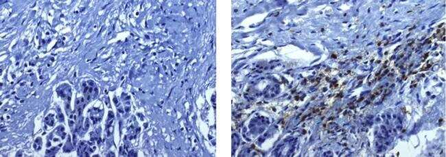
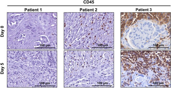
- Figure 6 CD45-positive lymphocytes remain viable for 5 days in human patient derived pancreatic ductal adenocarcinoma tumour explants. Immunohistochemistry was performed for lymphocyte marker CD45 on tumour explants from patients 1-3 at day 0 and day 5.
- Conjugate
- Biotin
- Submitted by
- Invitrogen Antibodies (provider)
- Main image
- Experimental details
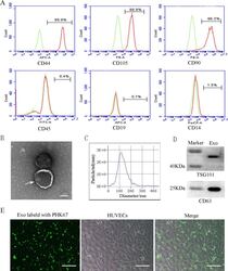
- Characterization and uptake of SHED-Exos. A Surface markers of SHED cells were analyzed by flow cytometry (FACS) and were positive for mesenchymal markers (CD44, CD105 and CD90) and negative for endothelial markers (CD45, CD19 and CD14). B The morphology of exosomes (indicated by arrows) was observed using a transmission electron microscope (TEM). Scale bar = 100 nm. C Particle size distribution of SHED-Exos assessed by nanoparticle tracking analysis (NTA). D Expression of exosome-specific CD63 and TSG101 validated using western blotting. E Efficient uptake of PKH67-labeled exosomes by HUVECs was detected at 24 h. Scale bars = 100 mum
- Conjugate
- Biotin
- Submitted by
- Invitrogen Antibodies (provider)
- Main image
- Experimental details
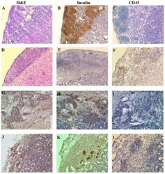
- Figure 9. Histology and immunohistochemical analysis of NICC xenografts. Representative hematoxylin and eosin staining images; and immunohistochemical staining images of porcine insulin and human CD45 in NICC xenograft samples from mice receiving (A-C) no human cells (NICC alone 84 days post-transplantation), (D-F) only human PBMCs (NICC + PBMC 28 days post-PBMC transfer), (G-I) human PBMCs and Xeno-Treg (NICC + PBMC + Xeno-Treg 84 days post-cell transfer) or (J-L) human PBMCs and Poly-Treg (NICC + PBMC + Poly-Treg 63 days post-cell transfer). (A, B, E-I, K and L) Magnification, x200; (C, D and J) magnification, x100. CD, cluster of differentiation; NICC, neonatal porcine islet cell clusters; PBMC, peripheral blood mononuclear cell; Poly-Treg, polyclonal Treg; Tregs, regulatory T cells; Xeno-Treg, Treg with xenoantigen specificity.
- Conjugate
- Biotin
