Explore
 Validate
 Learn
Learn Western blot
 Immunoprecipitation
Antibody data
- Antibody Data
- Antigen structure
- References [10]
- Comments [0]
- Validations
- Western blot [1]
- Other assay [4]
Submit
Validation data
Reference
Comment
Report error
- Product number
- AHO1011 - Provider product page

- Provider
- Invitrogen Antibodies
- Product name
- ErbB2 (HER-2) Monoclonal Antibody (e2-4001)
- Antibody type
- Monoclonal
- Antigen
- Other
- Reactivity
- Human, Mouse
- Host
- Mouse
- Isotype
- IgG
- Antibody clone number
- e2-4001
- Vial size
- 100 µg
- Concentration
- 1.0 mg/mL
- Storage
- -20°C
Submitted references SOX9/FXYD3/Src Axis Is Critical for ER(+) Breast Cancer Stem Cell Function.
HDAC5-mediated deacetylation and nuclear localisation of SOX9 is critical for tamoxifen resistance in breast cancer.
Molecular Correlates of In Vitro Responses to Dacomitinib and Afatinib in Bladder Cancer.
Integrated Molecular Characterization of Uterine Carcinosarcoma.
Context Specificity in Causal Signaling Networks Revealed by Phosphoprotein Profiling.
A Novel Affibody-Auristatin E Conjugate With a Potent and Selective Activity Against HER2+ Cell Lines.
Use of a genetically engineered mouse model as a preclinical tool for HER2 breast cancer.
Molecular architecture of the ErbB2 extracellular domain homodimer.
Pertuzumab counteracts the inhibitory effect of ErbB2 on degradation of ErbB3.
Glycerophospholipid profile in oncogene-induced senescence.
Xue Y, Lai L, Lian W, Tu X, Zhou J, Dong P, Su D, Wang X, Cao X, Chen Y, Wang Q
Molecular cancer research : MCR 2019 Jan;17(1):238-249
Molecular cancer research : MCR 2019 Jan;17(1):238-249
HDAC5-mediated deacetylation and nuclear localisation of SOX9 is critical for tamoxifen resistance in breast cancer.
Xue Y, Lian W, Zhi J, Yang W, Li Q, Guo X, Gao J, Qu H, Lin W, Li Z, Lai L, Wang Q
British journal of cancer 2019 Dec;121(12):1039-1049
British journal of cancer 2019 Dec;121(12):1039-1049
Molecular Correlates of In Vitro Responses to Dacomitinib and Afatinib in Bladder Cancer.
Tamura S, Wang Y, Veeneman B, Hovelson D, Bankhead A 3rd, Broses LJ, Lorenzatti Hiles G, Liebert M, Rubin JR, Day KC, Hussain M, Neamati N, Tomlins S, Palmbos PL, Grivas P, Day ML
Bladder cancer (Amsterdam, Netherlands) 2018 Jan 20;4(1):77-90
Bladder cancer (Amsterdam, Netherlands) 2018 Jan 20;4(1):77-90
Integrated Molecular Characterization of Uterine Carcinosarcoma.
Cherniack AD, Shen H, Walter V, Stewart C, Murray BA, Bowlby R, Hu X, Ling S, Soslow RA, Broaddus RR, Zuna RE, Robertson G, Laird PW, Kucherlapati R, Mills GB, Cancer Genome Atlas Research Network, Weinstein JN, Zhang J, Akbani R, Levine DA
Cancer cell 2017 Mar 13;31(3):411-423
Cancer cell 2017 Mar 13;31(3):411-423
Context Specificity in Causal Signaling Networks Revealed by Phosphoprotein Profiling.
Hill SM, Nesser NK, Johnson-Camacho K, Jeffress M, Johnson A, Boniface C, Spencer SE, Lu Y, Heiser LM, Lawrence Y, Pande NT, Korkola JE, Gray JW, Mills GB, Mukherjee S, Spellman PT
Cell systems 2017 Jan 25;4(1):73-83.e10
Cell systems 2017 Jan 25;4(1):73-83.e10
A Novel Affibody-Auristatin E Conjugate With a Potent and Selective Activity Against HER2+ Cell Lines.
Sochaj-Gregorczyk AM, Serwotka-Suszczak AM, Otlewski J
Journal of immunotherapy (Hagerstown, Md. : 1997) 2016 Jul-Aug;39(6):223-32
Journal of immunotherapy (Hagerstown, Md. : 1997) 2016 Jul-Aug;39(6):223-32
Use of a genetically engineered mouse model as a preclinical tool for HER2 breast cancer.
Creedon H, Balderstone LA, Muir M, Balla J, Gomez-Cuadrado L, Tracey N, Loane J, Klinowska T, Muller WJ, Brunton VG
Disease models & mechanisms 2016 Feb;9(2):131-40
Disease models & mechanisms 2016 Feb;9(2):131-40
Molecular architecture of the ErbB2 extracellular domain homodimer.
Hu S, Sun Y, Meng Y, Wang X, Yang W, Fu W, Guo H, Qian W, Hou S, Li B, Rao Z, Lou Z, Guo Y
Oncotarget 2015 Jan 30;6(3):1695-706
Oncotarget 2015 Jan 30;6(3):1695-706
Pertuzumab counteracts the inhibitory effect of ErbB2 on degradation of ErbB3.
Sak MM, Szymanska M, Bertelsen V, Hasmann M, Madshus IH, Stang E
Carcinogenesis 2013 Sep;34(9):2031-8
Carcinogenesis 2013 Sep;34(9):2031-8
Glycerophospholipid profile in oncogene-induced senescence.
Cadenas C, Vosbeck S, Hein EM, Hellwig B, Langer A, Hayen H, Franckenstein D, Büttner B, Hammad S, Marchan R, Hermes M, Selinski S, Rahnenführer J, Peksel B, Török Z, Vígh L, Hengstler JG
Biochimica et biophysica acta 2012 Sep;1821(9):1256-68
Biochimica et biophysica acta 2012 Sep;1821(9):1256-68
No comments: Submit comment
Supportive validation
- Submitted by
- Invitrogen Antibodies (provider)
- Main image
- Experimental details
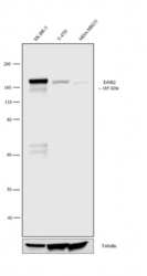
- Western blot analysis was performed on membrane enriched extracts (30 µg lysate) of SK-BR-3 (1), T-47D (2) and MDA-MB-231 (3). The blot was probed with anti-ErbB2 Mouse Monoclonal Antibody (Product # AHO1011, 1:500 dilution) and detected by chemiluminescence using Goat anti-Mouse IgG (H+L) Superclonal™ Secondary Antibody, HRP conjugate (Product # A28177, 0.25 µg/mL, 1:4000 dilution). A 185 kDa band corresponding to ErbB2 was observed in SK-BR-3 and moderate in T-47D where as it was not observed in MDA-MB-231 which is an ErbB2 negative cell line. Known quantity of protein samples were electrophoresed using Novex® NuPAGE® 4-12 % Bis-Tris gel (Product # NP0321BOX), XCell SureLock™ Electrophoresis System (Product # EI0002) and Novex® Sharp Pre-Stained Protein Standard (Product # LC5800). Resolved proteins were then transferred onto a nitrocellulose membrane with overnight wet transfer system. The membrane was probed with the relevant primary and secondary Antibody following blocking with 5 % skimmed milk. Chemiluminescent detection was performed using Pierce™ ECL Western Blotting Substrate (Product # 32106).

Supportive validation
- Submitted by
- Invitrogen Antibodies (provider)
- Main image
- Experimental details
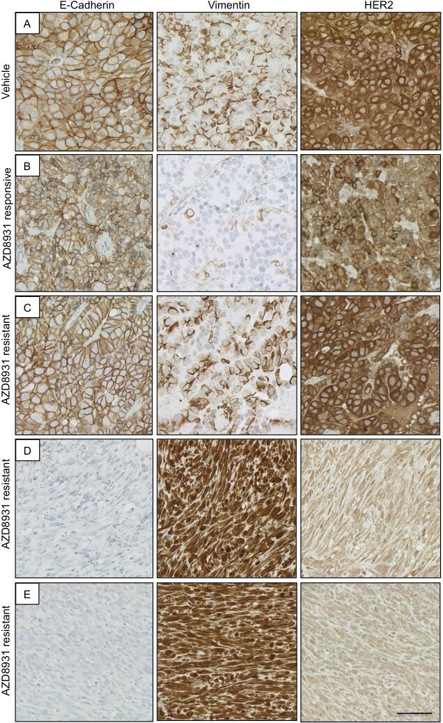
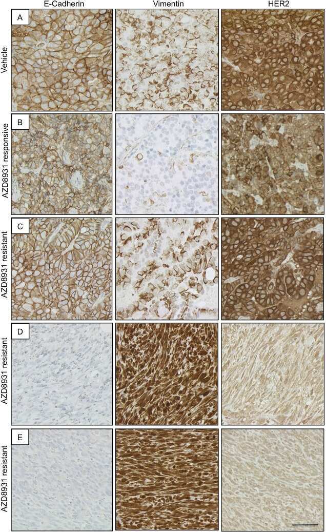
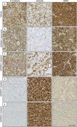
- Fig. 6. Development of EMT in AZD8931-resistant tumours. Immunohistochemical analysis of E-cadherin, vimentin and HER2 in (A) vehicle- and (B-E) AZD8931-treated tumours. (B) AZD8931-responsive tumour with growth inhibited by AZD8931. (C) AZD8931-resistant tumour that has retained the morphology of the AZD8931-naive (vehicle) tumours. (D,E) AZD8931-resistant tumours with a spindle cell morphology. Scale bar: 50 um.
- Submitted by
- Invitrogen Antibodies (provider)
- Main image
- Experimental details
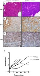
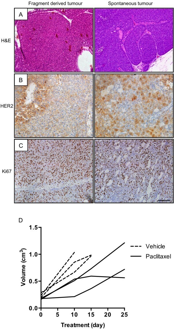
- Fig. 2. Transplantation of NIC-PTEN +/- fragments generated tumours that were indistinguishable from the parental tumours. (A) Representative H&E images of fragment-derived and spontaneous tumours from NIC-PTEN +/- mice. (B,C) Representative images of HER2 (B) and Ki67 (C) expression in fragment-derived and spontaneous tumours. Scale bar: 100 um. (D) Growth rate of vehicle- ( n =3) and paclitaxel-treated ( n =3) fragment-derived tumours.
- Submitted by
- Invitrogen Antibodies (provider)
- Main image
- Experimental details
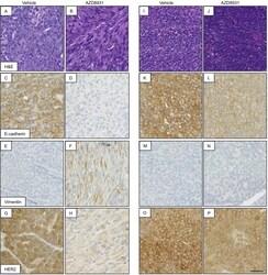
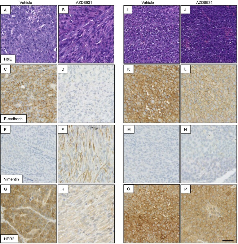
- Fig. 4. AZD8931 resistance is associated with EMT in a subpopulation of tumours. Analysis of AZD8931-naive (vehicle) and AZD8931-resistant tumours showing representative images of H&E staining and immunohistochemical analysis of E-cadherin, vimentin and HER2. Scale bar: 50 um. (A-H) AZD8931-resistant spindle cell tumour and corresponding vehicle-treated tumour. (I-P) AZD8931-resistant tumour phenotypically indistinguishable from AZD8931-naive (vehicle) tumour and corresponding vehicle-treated tumour.
- Submitted by
- Invitrogen Antibodies (provider)
- Main image
- Experimental details
- Fig. 6. Development of EMT in AZD8931-resistant tumours. Immunohistochemical analysis of E-cadherin, vimentin and HER2 in (A) vehicle- and (B-E) AZD8931-treated tumours. (B) AZD8931-responsive tumour with growth inhibited by AZD8931. (C) AZD8931-resistant tumour that has retained the morphology of the AZD8931-naive (vehicle) tumours. (D,E) AZD8931-resistant tumours with a spindle cell morphology. Scale bar: 50 um.