 Explore
 Validate
 Learn
Learn Immunocytochemistry
Antibody data
- Antibody Data
- Antigen structure
- References [24]
- Comments [0]
- Validations
- Immunocytochemistry [1]
- Immunohistochemistry [3]
- Other assay [8]
Submit
Validation data
Reference
Comment
Report error
- Product number
- PA5-16446 - Provider product page

- Provider
- Invitrogen Antibodies
- Product name
- Ki-67 Polyclonal Antibody
- Antibody type
- Polyclonal
- Antigen
- Synthetic peptide
- Description
- PA5-16446 targets Ki-67 in IHC (P) applications and shows reactivity with Rat and Human samples.
- Concentration
- 1 mg/mL
Submitted references Quercetin-Rich Extracts from Onions (Allium cepa) Play Potent Cytotoxicity on Adrenocortical Carcinoma Cell Lines, and Quercetin Induces Important Anticancer Properties.
MELK Inhibition Effectively Suppresses Growth of Glioblastoma and Cancer Stem-Like Cells by Blocking AKT and FOXM1 Pathways.
Curative effect of methotrexate combined with teniposide in the treatment of primary central nervous system lymphoma.
Boiling Histotripsy Ablation of Renal Cell Carcinoma in the Eker Rat Promotes a Systemic Inflammatory Response.
Mouse Nr2f1 haploinsufficiency unveils new pathological mechanisms of a human optic atrophy syndrome.
Interactive histogenesis of axonal strata and proliferative zones in the human fetal cerebral wall.
Knockout of MARCH2 inhibits the growth of HCT116 colon cancer cells by inducing endoplasmic reticulum stress.
Porf-2 Inhibits Neural Stem Cell Proliferation Through Wnt/β-Catenin Pathway by Its GAP Domain.
Ki-67 is overexpressed in human laryngeal carcinoma and contributes to the proliferation of HEp2 cells.
FAK regulates platelet extravasation and tumor growth after antiangiogenic therapy withdrawal.
Inhibition of STAT3 activity delays obesity-induced thyroid carcinogenesis in a mouse model.
Curcumin: a unique antioxidant offers a multimechanistic approach for management of hepatocellular carcinoma in rat model.
Zbed3 contributes to malignant phenotype of lung cancer via regulating β-catenin and P120-catenin 1.
Neoplastic transformation of porcine mammary epithelial cells in vitro and tumor formation in vivo.
Ductal activation of oncogenic KRAS alone induces sarcomatoid phenotype.
Oncogenic mutations of thyroid hormone receptor β.
The use of mouse models of breast cancer and quantitative image analysis to evaluate hormone receptor antigenicity after microwave-assisted formalin fixation.
CXCR4, but not CXCR7, discriminates metastatic behavior in non-small cell lung cancer cells.
Silencing MED1 sensitizes breast cancer cells to pure anti-estrogen fulvestrant in vitro and in vivo.
Dietary selenium deficiency exacerbates DSS-induced epithelial injury and AOM/DSS-induced tumorigenesis.
Oncogenic Actions of the Nuclear Receptor Corepressor (NCOR1) in a Mouse Model of Thyroid Cancer.
Reduced androgen receptor expression accelerates the onset of ERBB2 induced breast tumors in female mice.
Initiation of prostate cancer in mice by Tp53R270H: evidence for an alternative molecular progression.
Activation of neural and pluripotent stem cell signatures correlates with increased malignancy in human glioma.
Veiga AA, Irioda AC, Mogharbel BF, Bonatto SJR, Souza LM
Pharmaceuticals (Basel, Switzerland) 2022 Jun 16;15(6)
Pharmaceuticals (Basel, Switzerland) 2022 Jun 16;15(6)
MELK Inhibition Effectively Suppresses Growth of Glioblastoma and Cancer Stem-Like Cells by Blocking AKT and FOXM1 Pathways.
Zhang X, Wang J, Wang Y, Liu G, Li H, Yu J, Wu R, Liang J, Yu R, Liu X
Frontiers in oncology 2020;10:608082
Frontiers in oncology 2020;10:608082
Curative effect of methotrexate combined with teniposide in the treatment of primary central nervous system lymphoma.
Wang YX, Huang Y, Xu XP, Chen BB, Lin ZG, Ma Y, Ding TL, Wang Q
Oncology letters 2020 Mar;19(3):2097-2106
Oncology letters 2020 Mar;19(3):2097-2106
Boiling Histotripsy Ablation of Renal Cell Carcinoma in the Eker Rat Promotes a Systemic Inflammatory Response.
Schade GR, Wang YN, D'Andrea S, Hwang JH, Liles WC, Khokhlova TD
Ultrasound in medicine & biology 2019 Jan;45(1):137-147
Ultrasound in medicine & biology 2019 Jan;45(1):137-147
Mouse Nr2f1 haploinsufficiency unveils new pathological mechanisms of a human optic atrophy syndrome.
Bertacchi M, Gruart A, Kaimakis P, Allet C, Serra L, Giacobini P, Delgado-García JM, Bovolenta P, Studer M
EMBO molecular medicine 2019 Aug;11(8):e10291
EMBO molecular medicine 2019 Aug;11(8):e10291
Interactive histogenesis of axonal strata and proliferative zones in the human fetal cerebral wall.
Žunić Išasegi I, Radoš M, Krsnik Ž, Radoš M, Benjak V, Kostović I
Brain structure & function 2018 Dec;223(9):3919-3943
Brain structure & function 2018 Dec;223(9):3919-3943
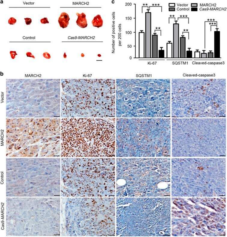
Knockout of MARCH2 inhibits the growth of HCT116 colon cancer cells by inducing endoplasmic reticulum stress.
Xia D, Ji W, Xu C, Lin X, Wang X, Xia Y, Lv P, Song Q, Ma D, Chen Y
Cell death & disease 2017 Jul 27;8(7):e2957
Cell death & disease 2017 Jul 27;8(7):e2957
Porf-2 Inhibits Neural Stem Cell Proliferation Through Wnt/β-Catenin Pathway by Its GAP Domain.
Huang GH, Yang XT, Chen K, Xing J, Guo L, Zhu L, Li HJ, Li XC, Zhang SY, Feng DF
Frontiers in cellular neuroscience 2016;10:85
Frontiers in cellular neuroscience 2016;10:85
Ki-67 is overexpressed in human laryngeal carcinoma and contributes to the proliferation of HEp2 cells.
Bai Y, Shao Y, Li H, Xue W, Quan F, Wu S
Oncology letters 2016 Oct;12(4):2641-2647
Oncology letters 2016 Oct;12(4):2641-2647
FAK regulates platelet extravasation and tumor growth after antiangiogenic therapy withdrawal.
Haemmerle M, Bottsford-Miller J, Pradeep S, Taylor ML, Choi HJ, Hansen JM, Dalton HJ, Stone RL, Cho MS, Nick AM, Nagaraja AS, Gutschner T, Gharpure KM, Mangala LS, Rupaimoole R, Han HD, Zand B, Armaiz-Pena GN, Wu SY, Pecot CV, Burns AR, Lopez-Berestein G, Afshar-Kharghan V, Sood AK
The Journal of clinical investigation 2016 May 2;126(5):1885-96
The Journal of clinical investigation 2016 May 2;126(5):1885-96
Inhibition of STAT3 activity delays obesity-induced thyroid carcinogenesis in a mouse model.
Park JW, Han CR, Zhao L, Willingham MC, Cheng SY
Endocrine-related cancer 2016 Jan;23(1):53-63
Endocrine-related cancer 2016 Jan;23(1):53-63
Curcumin: a unique antioxidant offers a multimechanistic approach for management of hepatocellular carcinoma in rat model.
Ahmed HH, Shousha WG, Shalby AB, El-Mezayen HA, Ismaiel NN, Mahmoud NS
Tumour biology : the journal of the International Society for Oncodevelopmental Biology and Medicine 2015 Mar;36(3):1667-78
Tumour biology : the journal of the International Society for Oncodevelopmental Biology and Medicine 2015 Mar;36(3):1667-78
Zbed3 contributes to malignant phenotype of lung cancer via regulating β-catenin and P120-catenin 1.
Fan C, Jiang G, Zhang X, Miao Y, Lin X, Luan L, Xu Z, Zhang Y, Zhao H, Liu D, Wang E
Molecular carcinogenesis 2015 Jun;54 Suppl 1:E138-47
Molecular carcinogenesis 2015 Jun;54 Suppl 1:E138-47
Neoplastic transformation of porcine mammary epithelial cells in vitro and tumor formation in vivo.
Rowson-Hodel AR, Manjarin R, Trott JF, Cardiff RD, Borowsky AD, Hovey RC
BMC cancer 2015 Jul 31;15:562
BMC cancer 2015 Jul 31;15:562
Ductal activation of oncogenic KRAS alone induces sarcomatoid phenotype.
Fu Y, Cruz-Monserrate Z, Helen Lin H, Chung Y, Ji B, Lin SM, Vonderfecht S, Logsdon CD, Li CF, Ann DK
Scientific reports 2015 Aug 20;5:13347
Scientific reports 2015 Aug 20;5:13347
Oncogenic mutations of thyroid hormone receptor β.
Park JW, Zhao L, Willingham M, Cheng SY
Oncotarget 2015 Apr 10;6(10):8115-31
Oncotarget 2015 Apr 10;6(10):8115-31
The use of mouse models of breast cancer and quantitative image analysis to evaluate hormone receptor antigenicity after microwave-assisted formalin fixation.
Engelberg JA, Giberson RT, Young LJ, Hubbard NE, Cardiff RD
The journal of histochemistry and cytochemistry : official journal of the Histochemistry Society 2014 May;62(5):319-34
The journal of histochemistry and cytochemistry : official journal of the Histochemistry Society 2014 May;62(5):319-34
CXCR4, but not CXCR7, discriminates metastatic behavior in non-small cell lung cancer cells.
Choi YH, Burdick MD, Strieter BA, Mehrad B, Strieter RM
Molecular cancer research : MCR 2014 Jan;12(1):38-47
Molecular cancer research : MCR 2014 Jan;12(1):38-47
Silencing MED1 sensitizes breast cancer cells to pure anti-estrogen fulvestrant in vitro and in vivo.
Zhang L, Cui J, Leonard M, Nephew K, Li Y, Zhang X
PloS one 2013;8(7):e70641
PloS one 2013;8(7):e70641
Dietary selenium deficiency exacerbates DSS-induced epithelial injury and AOM/DSS-induced tumorigenesis.
Barrett CW, Singh K, Motley AK, Lintel MK, Matafonova E, Bradley AM, Ning W, Poindexter SV, Parang B, Reddy VK, Chaturvedi R, Fingleton BM, Washington MK, Wilson KT, Davies SS, Hill KE, Burk RF, Williams CS
PloS one 2013;8(7):e67845
PloS one 2013;8(7):e67845
Oncogenic Actions of the Nuclear Receptor Corepressor (NCOR1) in a Mouse Model of Thyroid Cancer.
Fozzatti L, Park JW, Zhao L, Willingham MC, Cheng SY
PloS one 2013;8(6):e67954
PloS one 2013;8(6):e67954
Reduced androgen receptor expression accelerates the onset of ERBB2 induced breast tumors in female mice.
Hodgson MC, Vanostran G, Alghamdi S, Poppiti RJ, Agoulnik AI, Agoulnik IU
PloS one 2013;8(4):e60455
PloS one 2013;8(4):e60455
Initiation of prostate cancer in mice by Tp53R270H: evidence for an alternative molecular progression.
Vinall RL, Chen JQ, Hubbard NE, Sulaimon SS, Shen MM, Devere White RW, Borowsky AD
Disease models & mechanisms 2012 Nov;5(6):914-20
Disease models & mechanisms 2012 Nov;5(6):914-20
Activation of neural and pluripotent stem cell signatures correlates with increased malignancy in human glioma.
Holmberg J, He X, Peredo I, Orrego A, Hesselager G, Ericsson C, Hovatta O, Oba-Shinjo SM, Marie SK, Nistér M, Muhr J
PloS one 2011 Mar 31;6(3):e18454
PloS one 2011 Mar 31;6(3):e18454
No comments: Submit comment
Supportive validation
- Submitted by
- Invitrogen Antibodies (provider)
- Main image
- Experimental details
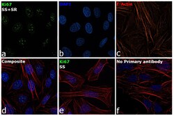
- Immunofluorescence analysis of Ki67 was performed using 70% confluent log phase HeLa cells serum starved for 36 Hrs followed by serum release for 6 Hrs. The cells were fixed with 4% Paraformaldehyde for 10 minutes, permeabilized with 0.1% Triton™ X-100 for 10 minutes, and blocked with 2% BSA for 10 minutes at room temperature. The cells were labeled with Ki-67 Polyclonal Antibody (Product # PA5-16446) at 1:100 dilution in 0.1% BSA, incubated at 4 degree celsius overnight and then labeled with Goat anti-Rabbit IgG (H+L) Superclonal™ Secondary Antibody, Alexa Fluor® 488 conjugate (Product # A27034, 1:2000 dilution) for 45 minutes at room temperature (Panel a: Green). Nuclei (Panel b: Blue) were stained with SlowFade® Gold Antifade Mountant with DAPI (Product # S36938). F-actin (Panel c: Red) was stained with Rhodamine Phalloidin (Product # R415, 1:300). Panel d represents the merged image showing nuclear localization. Panel e represents serum starved cells with reduced signal. Panel f represents control cells with no primary antibody to assess background. The images were captured at 60X magnification.
Supportive validation
- Submitted by
- Invitrogen Antibodies (provider)
- Main image
- Experimental details
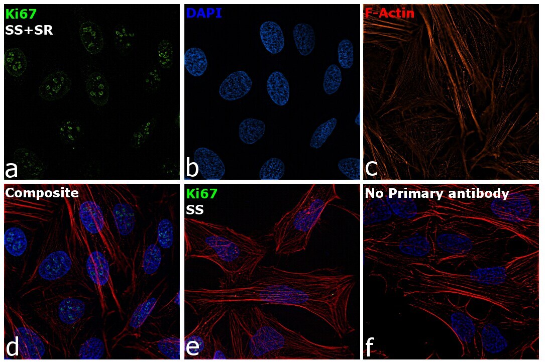
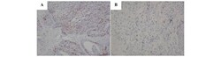
- Formalin-fixed, paraffin-embedded human tonsil stained with Ki67 antibody using peroxidase-conjugate and AEC chromogen. Note nuclear staining of proliferating cells.

- Submitted by
- Invitrogen Antibodies (provider)
- Main image

- Experimental details
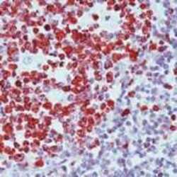
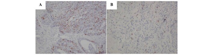
- Immunohistochemistry analysis of Ki-67 showing staining in the nucleus of paraffin-embedded human breast tissue (right) compared to a negative control without primary antibody (left). To expose target proteins, antigen retrieval was performed using 10mM sodium citrate (pH 6.0), microwaved for 8-15 min. Following antigen retrieval, tissues were blocked in 3% H2O2-methanol for 15 min at room temperature, washed with ddH2O and PBS, and then probed with a Ki-67 Rabbit Polyclonal Antibody (Product # PA5-16446) diluted in 3% BSA-PBS at a dilution of 1:20 for 1 hour at 37°C in a humidified chamber. Tissues were washed extensively in PBST and detection was performed using an HRP-conjugated secondary antibody followed by colorimetric detection using a DAB kit. Tissues were counterstained with hematoxylin and dehydrated with ethanol and xylene to prep for mounting.
- Submitted by
- Invitrogen Antibodies (provider)
- Main image

- Experimental details
- Immunohistochemistry analysis of Ki-67 showing staining in the nucleus of paraffin-embedded human tonsil tissue (right) compared to a negative control without primary antibody (left). To expose target proteins, antigen retrieval was performed using 10mM sodium citrate (pH 6.0), microwaved for 8-15 min. Following antigen retrieval, tissues were blocked in 3% H2O2-methanol for 15 min at room temperature, washed with ddH2O and PBS, and then probed with a Ki-67 Rabbit Polyclonal Antibody (Product # PA5-16446) diluted in 3% BSA-PBS at a dilution of 1:20 for 1 hour at 37°C in a humidified chamber. Tissues were washed extensively in PBST and detection was performed using an HRP-conjugated secondary antibody followed by colorimetric detection using a DAB kit. Tissues were counterstained with hematoxylin and dehydrated with ethanol and xylene to prep for mounting.

Supportive validation
- Submitted by
- Invitrogen Antibodies (provider)
- Main image

- Experimental details
- NULL
- Submitted by
- Invitrogen Antibodies (provider)
- Main image

- Experimental details
- NULL
- Submitted by
- Invitrogen Antibodies (provider)
- Main image
- Experimental details
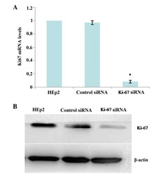
- Figure 4. Knockdown of Ki-67 expression in HEp2 cells at 24 h after siRNA transfection. (A) Ki-67 mRNA levels were determined by RT-qPCR. Relative fold induction for Ki-67 mRNA (means +- SD) in the mock- and Ki-67 siRNA-transfected HEp2 cells is presented relative to the expression in the parental HEp2 cells (*P

- Submitted by
- Invitrogen Antibodies (provider)
- Main image

- Experimental details
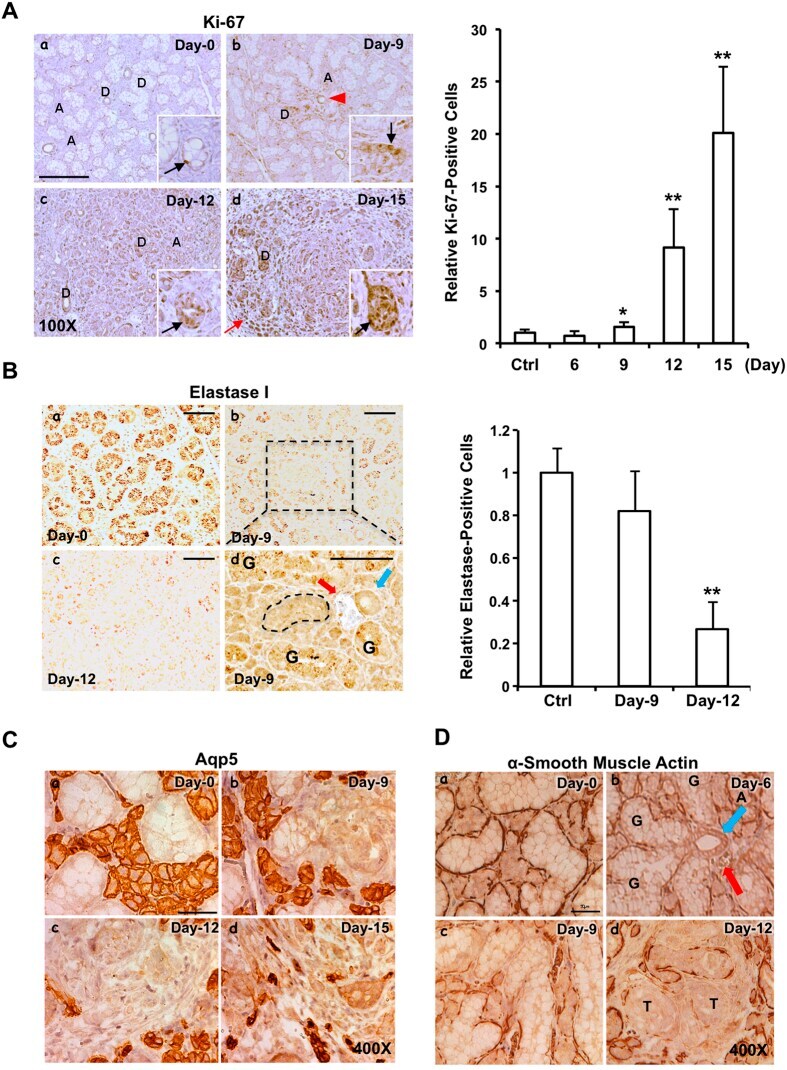
- Figure 4 Cell proliferation starting from day-9 following oncogenic KRAS activation. ( A ) Nuclear Ki-67-positive cells increased mainly at the ductal cells starting from day-9 post KRAS G12V induction. Scale bars: 250 mum; inset : 200X; A: acini; D: duct; black arrow: nuclear Ki-67-positive ductal cell; red arrow: nuclear Ki-67-positive fibroblast; red arrowhead: blood vessel. The relative fold change in the nuclear Ki-67-positive staining with the level in the SMGs of LGL-KRas G12V ;Ela-CreERT mice prior to Tam-feeding (Ctrl) set as 1. Results are shown as mean +- S.D. enumerated from 6 randomly selected non-overlapping fields (10X) by Image-Pro Premier 9.0; * p < 0.05, ** p < 0.01. ( B ) Elastase I accumulation in the SMG ductal cells of LGL-KRas G12V ;Ela-CreERT mice decreased during the course of tumorigenesis ( left panels ). Panel d (enlarged from boxed region in panel b ): elastase I staining was lost in a GCT (shown by dotted line, panel d ) near a dyad structure of a striated duct (blue arrow) and a blood vessel (red arrow); residual elastase I staining was visualized in some GCTs (label with G). Scale bar: 100 mum ( panels a-c :); 100 mum ( panel d , enlarged from b ). The relative fold change in the elastase I-positive staining ( right panel ). Quantitative analyses of elastase I-positive staining with level at control SMGs set as 1. Results are shown as mean +- S.D. enumerated from 5 randomly selected non-overlapping fields (10X) by Image-Pro Premier 9.0; ** p
- Submitted by
- Invitrogen Antibodies (provider)
- Main image
- Experimental details
- Figure 1. Immunohistochemical staining of Ki-67 protein of laryngeal squamous carcinoma tissues and the corresponding non-cancerous tissues (magnification, x200). (A) Strong immunostaining for Ki-67 protein in laryngeal squamous carcinoma tissue. (B) Ki-67 protein expression is absent in non-cancerous laryngeal tissue.
- Submitted by
- Invitrogen Antibodies (provider)
- Main image

- Experimental details
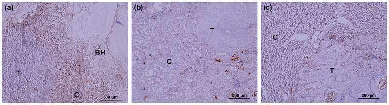
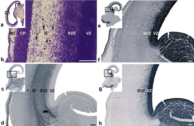
- Fig. 1 Early sagittal fibers on coronal Nissl ( a, b ) and immunostained histological sections ( c - h ) through the occipital ( a, b ) and midlateral levels ( c - h ) of the brain at 9 ( a, b ) and 11 ( c - h ) PCW. The rectangle in 'a', 'c', 'e', and 'g' are shown at higher magnifications in 'b', 'd', 'f', and 'h' respectively. The intermediate zone (IZ) contains large fiber bundles ( b asterix), running in a sagittal direction separated by groups of migratory neurons ( b arrowheads). Coronal sections through the brain at 11 PCW showed SNAP25 immuno-positivity to sagittal fibers in the IZ ( c, d ). Some fibers originating from the thalamus showed radial orientation before attaining sagittal orientation ( d arrowhead). The IZ is well delineated from the presubplate ( d asterisk) and ventricular zone (VZ), but intermingles with the subventricular zone (SVZ). Staining of the same brain (11 PCW) for SOX2 ( e, f ) and Ki67 ( g, h ) showing a homogenous VZ but dispersed SVZ. ( CP cortical plate; MZ marginal zone). Scale bar = 100 um ( b ); 200 um ( d, f )
- Submitted by
- Invitrogen Antibodies (provider)
- Main image

- Experimental details
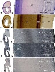
- Fig. 12 Maturation of the occipital sagittal strata (SS) on coronal plane T2 MR images ( a, h ), and corresponding histological preparations from newborn ( b - g ) and preterm ( i - n ) brains. The rectangle in b, d, f, i, k , and m are shown at higher magnification in c, e, g, j, l , and n , respectively. Comparison of MRI scans through the occipital lobe in a newborn infant ( a ) and a premature infant scanned at term equivalent age ( h ) suggested a slight difference in visibility and delineation of the sagittal strata ( a , h arrow) fashioned as a visible ""triplet structure"". Histological sections of Nissl-stained ( b, c; i, j ) preparations, sections immunostained for the proliferative marker Ki67 ( d, e; k, l ) and myelin basic protein SMI99 ( f, g; m, n ) indicated that different underlying factors contribute to the MRI ""triplet"" appearance in preterm and term periods. The differential prominence of cell and fiber layers, and the differential appearance of proliferative cells and myelination of the visual pathway at preterm and term determined the structure of the occipital SS. The less visible ""triplet structure"" in a preterm infant at TEA ( h ) was probably due to the disappearance of the transient proliferative cell band (arrow, l ; please, see MRI scan of preterm brain scanned soon after birth on Fig. 9 ) and impaired myelination ( n arrow). In the newborn brain, histological coronal sections through the occipital lobe ( f, g ), at the level of the

- Submitted by
- Invitrogen Antibodies (provider)
- Main image
- Experimental details
- Fig. 8 Laminar organization of the sagittal strata during the early preterm period. Coronal Nissl-stained frozen section ( a, b ) through the parietooccipital portion of the brain at 26 PCW showing a similar laminar organization as seen in the previous phases (see Fig. 5 ). The internal sagittal stratum (ISS) is densely cellular ( b number 5), and this stratum may be partially defined on both AChE-stained and unstained portion ( d ) on section of the brain at the same age. At this age, both cellular (O)SVZ and former fibrillar intermediate zone now are incorporated in multilaminar axonal-cellular compartment (MACC). Rectangles in a, c, e, g , and i are shown at higher magnifications in b, d, f, h , and j , respectively. Sublamination of the sagittal fiber strata and related cell layers were also visible on horizontal histological sections of brain at 24.5 PCW when immunostained for vimentin ( e, f ), GFAP ( g, h ), and Ki67 ( i, j ). Numbers (1-8) used to mark the laminas are explained in detail in Fig. 5 . Immunostaining for glial markers ( e - h ) showed that the radial glia transverses the radial-sagittal strata and subplate (SP) to reach the cortical plate (CP). Immunostaining for proliferative markers ( i, j ) showed proliferative activity in cellular-fibrillar layers, except in the compact periventricular (callosal) system. The two proliferative cell layers ( b between arrows) were not a constant finding on Nissl-stained preparations of the occipital lobe (see
