 Explore
 Validate
 Learn
Learn Western blot
Antibody data
- Antibody Data
- Antigen structure
- References [1]
- Comments [0]
- Validations
- Western blot [4]
- Immunocytochemistry [1]
- Other assay [1]
Submit
Validation data
Reference
Comment
Report error
- Product number
- 702187 - Provider product page

- Provider
- Invitrogen Antibodies
- Product name
- B-Raf Recombinant Rabbit Monoclonal Antibody (7H30L21)
- Antibody type
- Monoclonal
- Antigen
- Other
- Description
- This antibody is predicted to react with Monkey, Sheep, Dog and Mouse
- Antibody clone number
- 7H30L21
- Concentration
- 0.5 mg/mL
Submitted references Inducible Degradation of Target Proteins through a Tractable Affinity-Directed Protein Missile System.
Simpson LM, Macartney TJ, Nardin A, Fulcher LJ, Röth S, Testa A, Maniaci C, Ciulli A, Ganley IG, Sapkota GP
Cell chemical biology 2020 Sep 17;27(9):1164-1180.e5
Cell chemical biology 2020 Sep 17;27(9):1164-1180.e5
No comments: Submit comment
Supportive validation
- Submitted by
- Invitrogen Antibodies (provider)
- Main image

- Experimental details
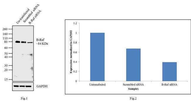
- Knockdown of B-Raf was achieved by transfecting NIH/3T3 cells with B-Raf specific validated siRNAs (Silencer® select Product # s2080, s2081, s2082). Western blot analysis (Fig a) was performed using membrane enriched extracts from the B-Raf knockdown cells (lane 3), non-specific scrambled siRNA transfected cells (lane 2) and untransfected cells (lane 1). The blots were probed with Anti-B-Raf Antibody, Recombinant Rabbit Monoclonal (Product # 702187, 1 µg/mL) and Goat anti-Rabbit IgG (H+L) Superclonal™ Secondary Antibody, HRP conjugate (Product # A27036, 0.25 µg/mL, 1:4000 dilution). Densitometric analysis of this Western blot is shown in histogram (Fig b). Decrease in signal upon siRNA mediated knock down confirms that antibody is specific to B-Raf.
- Submitted by
- Invitrogen Antibodies (provider)
- Main image
- Experimental details
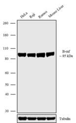
- Western blot analysis was performed on Whole cell extracts (30 µg lysate) of HeLa (Lane 1), Raji (Lane 2), Ramos (Lane 3) and tissue extracts of Mouse Liver (Lane 4). The blots were probed with Anti-B-raf Recombinant Rabbit Monoclonal Antibody (Product # 702187, 1-2 µg/mL) and detected by chemiluminescence using Goat anti-Rabbit IgG (H+L) Superclonal Secondary Antibody, HRP conjugate (Product # A27036, 0.4 µg/mL, 1:2500 dilution). A 85 kDa band corresponding to B-raf was observed across the cell lines and tissues tested. Known quantity of protein samples were electrophoresed using Novex®NuPAGE®4-12% Bis-Tris gel (Product # NP0321BOX), XCell SureLock Electrophoresis System (Product # EI0002) and Novex® Sharp Pre-Stained Protein Standard (Product # LC5800). Resolved proteins were then transferred onto a nitrocellulose membrane with iBlot® Dry Blotting System (Product # IB21001). The membrane was probed with the relevant primary and secondary Antibody following blocking with 5% skimmed milk. Chemiluminescent detection was performed using Pierce™ ECL Western blotting Substrate (Product # 32106).

- Submitted by
- Invitrogen Antibodies (provider)
- Main image

- Experimental details
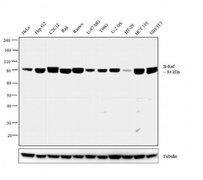
- Western blot analysis was performed on membrane enriched extracts (30 µg lysate) of HeLa (Lane 1), Hep G2 (Lane 2), C2C12 (Lane 3), Raji (Lane 4), Ramos (Lane 5), U-87 MG (Lane 6), T98G (Lane 7), U-2 OS (Lane 8), HT-29 (Lane 9), HCT116 (Lane 10) and NIH/3T3 (Lane 11). The blot was probed with Rabbit Anti-B-Raf Antibody, Recombinant Rabbit Monoclonal (Product # 702187, 1 µg/mL) and detected by chemiluminescence using Goat anti-Rabbit IgG (H+L) Superclonal™ Secondary Antibody, HRP conjugate (Product # A27036, 0.25 µg/mL, 1:4000 dilution). A 84 kDa band corresponding to B-Raf was observed across the cell lines tested. Known quantity of protein samples were electrophoresed using Novex®NuPAGE®4-12 % Bis-Tris gel (Product # NP0321BOX), XCell SureLock™ Electrophoresis System (Product # EI0002) and Novex® Sharp Pre-Stained Protein Standard (Product # LC5800). Resolved proteins were then transferred onto a nitrocellulose membrane with iBlot® 2 Dry Blotting System (Product # IB21001). The membrane was probed with the relevant primary and secondary Antibody following blocking with 5 % skimmed milk. Chemiluminescent detection was performed using Pierce™ ECL Western blotting Substrate (Product # 32106).
- Submitted by
- Invitrogen Antibodies (provider)
- Main image
- Experimental details
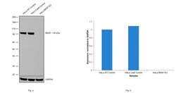
- Knockout of BRAF was achieved by CRISPR-Cas9 genome editing using LentiArray™ Lentiviral sgRNA (Product # A32042) (Assay ID CRISPR868710_LV) and LentiArray Cas9 Lentivirus (Product # A32064). Western blot analysis of BRAF was performed by loading 30 µg of HeLa wild type (Lane 1), HeLa CAS9 (Lane 2), HeLa BRAF KO (Lane 3) membrane extracts. The blot was probed with Anti-B-Raf Recombinant Rabbit Monoclonal Antibody (7H30L21)(Product # 702187) using 1 µg/mL dilution and Goat anti-Rabbit IgG (H+L), Superclonal™ Recombinant Secondary Antibody, HRP (Product # A27036). Loss of signal upon CRISPR mediated knockout (KO) using the LentiArray™ CRISPR product line confirms that antibody is specific to BRAF.

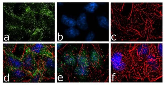
Supportive validation
- Submitted by
- Invitrogen Antibodies (provider)
- Main image

- Experimental details
- For immunofluorescence analysis, HeLa cells were fixed and permeabilized for detection of Braf using Anti-Braf Recombinant Rabbit Monoclonal Antibody (Product # 702187, 2 µg/mL) and labeled with Goat anti-Rabbit IgG (H+L) Superclonal Secondary Antibody, Alexa Fluor® 488 conjugate (Product # A27034, 1:2000). Panel a) shows representative cells that were stained for detection and localization of Braf protein (green), Panel b) is stained for nuclei (blue) using SlowFade® Gold Antifade Mountant with DAPI (Product # S36938). Panel c) represents cytoskeletal F-actin staining using Alexa Fluor® 555 Rhodamine Phalloidin (Product # R415, 1:300). Panel d) is a composite image of Panels a, b and c clearly demonstrating cytoplasmic localization of Braf. Panel e) shows translocation of Braf into nucleus upon EGF treatment (100 ng/mL 5 minutes). Panel f) represents control cells with no primary antibody to assess background. The images were captured at 60X magnification.
Supportive validation
- Submitted by
- Invitrogen Antibodies (provider)
- Main image

- Experimental details
- Figure 7 Untagged Endogenous RAS Proteins Are Degraded with HaloPROTAC-E in Cells Expressing FLAG-Halo-aHRAS (A) Schematic representation of FLAG-Halo-aHRAS HaloPROTAC L-AdPROM system. (B) A549 FLAG-empty, FLAG-aHRAS, and FLAG-Halo-aHRAS-expressing cells were lysed and subjected to IP with anti-FLAG M2 resin. F.T., post-IP flow-through extract. (C) A549 FLAG-Halo-aHRAS-expressing cells were treated with increasing concentrations of HaloPROTAC-E (0-10 muM) for 24 h. (D) A549 FLAG-Halo-aHRAS-expressing cells were treated with 500 nM HaloPROTAC-E for indicated times (0-48 h). (E) A549 FLAG-Halo-aHRAS-expressing cells were treated with 500 nM HaloPROTAC-E and 20 muM proteasome inhibitor MG132 for 24 h. (F) A549 FLAG-aHRAS and FLAG-Halo-aHRAS-expressing cells were treated with 500 nM HaloPROTAC-E for indicated times (0, 3, 6, and 24 h). For (B-F), extracts and IPs were resolved by SDS-PAGE and transferred on to PVDF membranes, which were subjected to immunoblotting with indicated antibodies. (G-K) Quantification from (F) of relative (G) panRAS normalized to GAPDH protein levels (n = 6 +- SD), (H) BRAF normalized to GAPDH protein levels (n = 3 +- SD), (I) p-Y1068 EGFR normalized to total EGFR protein levels (n = 6 +- SD), (J) p-T202/Y204 ERK1/2 normalized to total ERK1/2 protein levels (n = 6 +- SD), and (K) p-S473 Akt normalized to total Akt protein levels (n = 6 +- SD) in the absence or presence of HaloPROTAC-E (500 nM, 24 h). Statistical analyses were carried out by one-way anal
