 Explore
 Validate
 Learn
Learn Western blot
Antibody data
- Antibody Data
- Antigen structure
- References [2]
- Comments [0]
- Validations
- Western blot [2]
- Immunocytochemistry [1]
Submit
Validation data
Reference
Comment
Report error
- Product number
- 701510 - Provider product page

- Provider
- Invitrogen Antibodies
- Product name
- SQSTM1 Recombinant Rabbit Monoclonal Antibody (11HC14LC25)
- Antibody type
- Monoclonal
- Antigen
- Other
- Description
- This antibody is predicted to react with bovine, feline, non-human primate, mouse, rat, ovine and porcine based on sequence homology.
- Antibody clone number
- 11HC14LC25
- Concentration
- 0.5 mg/mL
Submitted references The pneumococcal two-component system SirRH is linked to enhanced intracellular survival of Streptococcus pneumoniae in influenza-infected pulmonary cells.
Composition of the Intranuclear Inclusions of Fragile X-associated Tremor/Ataxia Syndrome.
Reinoso-Vizcaíno NM, Cian MB, Cortes PR, Olivero NB, Hernandez-Morfa M, Piñas GE, Badapanda C, Rathore A, Perez DR, Echenique J
PLoS pathogens 2020 Aug;16(8):e1008761
PLoS pathogens 2020 Aug;16(8):e1008761
Composition of the Intranuclear Inclusions of Fragile X-associated Tremor/Ataxia Syndrome.
Ma L, Herren AW, Espinal G, Randol J, McLaughlin B, Martinez-Cerdeño V, Pessah IN, Hagerman RJ, Hagerman PJ
Acta neuropathologica communications 2019 Sep 3;7(1):143
Acta neuropathologica communications 2019 Sep 3;7(1):143
No comments: Submit comment
Supportive validation
- Submitted by
- Invitrogen Antibodies (provider)
- Main image

- Experimental details
- Western blot analysis of Sequestosome-1 recombinant protein (lane 1, 200 ng) and endogenous Sequestome-1 in extracts from HeLa cells treated with 60 mM chloroquinone for 16 hours (lane 2) using a Sequestome-1 Recombinant Rabbit Polyclonal Antibody (Product # 701510) at a concentration of 1 µg/mL. A rabbit Anti-tubulin antibody was blotted as a loading control and the blots were developed using chemiluminescence (ECL) method with a Goat anti-Rabbit HRP-conjugated secondary antibody (Product # G-21234). Results show a band at ~62 kDa.

- Submitted by
- Invitrogen Antibodies (provider)
- Main image
- Experimental details
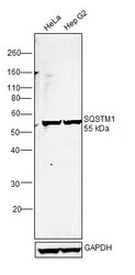
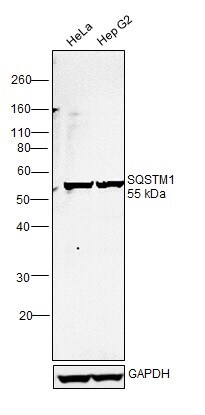
- Western blot was performed using Anti-SQSTM1 Recombinant Rabbit Monoclonal Antibody (11HC14LC25) (Product # 701510) and a ~55kDa band corresponding to SQSTM1 was observed across cell lines tested. Whole cell extracts (30 µg lysate) of HeLa (Lane 1) and Hep G2 (Lane 2) were electrophoresed using NuPAGE™ 10% Bis-Tris Protein Gel (Product # NP0301BOX). Resolved proteins were then transferred onto a Nitrocellulose membrane (Product # IB23001) by iBlot® 2 Dry Blotting System (Product # IB21001). The blot was probed with the primary antibody (0.5 µg/mL) and detected by chemiluminescence with Goat anti-Rabbit IgG (H+L) Superclonal™ Recombinant Secondary Antibody, HRP (Product # A27036, 1:4000 dilution using the iBright FL 1000 (Product # A32752). Chemiluminescent detection was performed using Novex® ECL Chemiluminescent Substrate Reagent Kit (Product # WP20005).
Supportive validation
- Submitted by
- Invitrogen Antibodies (provider)
- Main image
- Experimental details
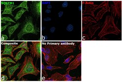
- Immunofluorescence analysis of SQSTM1 was performed using 70% confluent log phase HeLa cells. The cells were fixed with 4% paraformaldehyde for 10 minutes, permeabilized with 0.1% Triton™ X-100 for 15 minutes, and blocked with 2% BSA for 45 minutes at room temperature. The cells were labeled with SQSTM1 Recombinant Rabbit Monoclonal Antibody (11HC14LC25) (Product # 701510) at 1 µg/mL in 0.1% BSA, incubated at 4 degree celsius overnight and then labeled with Goat anti-Rabbit IgG (H+L) Highly Cross-Adsorbed Secondary Antibody, Alexa Fluor Plus 488 (Product # A32731), (1:2000 dilution), for 45 minutes at room temperature (Panel a: Green). Nuclei (Panel b:Blue) were stained with ProLong™ Diamond Antifade Mountant with DAPI (Product # P36962). F-actin (Panel c: Red) was stained with Rhodamine Phalloidin (Product # R415, 1:300 dilution). Panel d represents the merged image showing cytoplasm, endosome-like, plasma membrane and nuclear localization. Panel e represents control cells with no primary antibody to assess background. The images were captured at 60X magnification.
