Explore
 Validate
 Learn
LearnMIA1101
antibody from Invitrogen Antibodies
Targeting: SERPINA1
A1A, A1AT, AAT, alpha-1-antitrypsin, alpha1AT, PI, PI1
 Western blot
Antibody data
- Antibody Data
- Antigen structure
- References [0]
- Comments [0]
- Validations
- Western blot [3]
- Immunocytochemistry [1]
Submit
Validation data
Reference
Comment
Report error
- Product number
- MIA1101 - Provider product page

- Provider
- Invitrogen Antibodies
- Product name
- alpha-1 Antitrypsin Monoclonal Antibody (TMF1#4B5)
- Antibody type
- Monoclonal
- Antigen
- Purifed from natural sources
- Description
- MIA1101 targets Alpha1-Antitrypsin in IP applications and shows reactivity with Human samples.
- Antibody clone number
- TMF1#4B5
- Concentration
- 5 mg/mL
No comments: Submit comment
Supportive validation
- Submitted by
- Invitrogen Antibodies (provider)
- Main image

- Experimental details
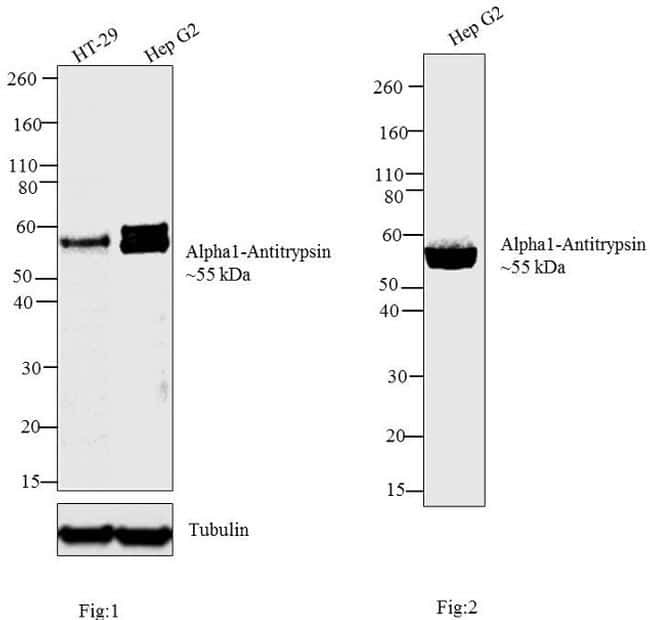
- Western blot analysis was performed on membrane enriched extracts (30 µg) of (Fig 1) HT-29 (Lane 1) and Hep G2 (Lane 2). Likewise, Western blot analysis was performed on (Fig 2) condition media of Hep G2 (Lane 1).The blots were probed with Anti-Alpha1-Antitrypsin Mouse Monoclonal Antibody (Product # MIA1101, 2 µg/mL) and detected by chemiluminescence using Goat anti-Mouse IgG (H+L) Superclonal™ Secondary Antibody, HRP conjugate (Product # A28177, 0.4 µg/mL, 1:2500 dilution).A~55 kDa band corresponding Alpha1-Antitrypsin was observed in HT-29, Hep G2 and in Hep G2 conditioned media. The observed shift in molecular weight from 50 kDa is due to glycosylation of the protein. Known quantity of protein samples were electrophoresed using Novex® NuPAGE®4-12 % Bis-Tris gel (Product # NP0321BOX), XCell SureLock™ Electrophoresis System (Product # EI0002) and Novex® Sharp Pre-Stained Protein Standard (Product # LC5800). Resolved proteins were then transferred onto a nitrocellulose membrane by iBlot® 2 Dry Blotting System (Product # IB21001). The membrane was probed with the relevant primary and secondary Antibody using iBind™ Flex Western Starter Kit (Product # SLF2000S). Chemiluminescent detection was performed using Pierce™ ECL Western Blotting Substrate (Product # 32106).
- Submitted by
- Invitrogen Antibodies (provider)
- Main image
- Experimental details
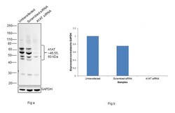
- Knockdown of alpha-1 Antitrypsin was achieved by transfecting Hep G2 with alpha-1 Antitrypsin specific siRNAs (Silencer® select Product # S10459, S10458). Western blot analysis (Fig. a) was performed using Membrane enriched extracts from the alpha-1 Antitrypsin knockdown cells (lane 3), non-targeting scrambled siRNA transfected cells (lane 2) and untransfected cells (lane 1). The blot was probed with alpha-1 Antitrypsin Monoclonal Antibody (TMF1#4B5) (Product # MIA1101, 2 µg/mL and Goat anti-Mouse IgG (H+L) Superclonal™ Recombinant Secondary Antibody, HRP (Product # A28177, 1:4000 dilution). Densitometric analysis of this western blot is shown in histogram (Fig. b). Decrease in signal upon siRNA mediated knock down confirms that antibody is specific to alpha-1 Antitrypsin.

- Submitted by
- Invitrogen Antibodies (provider)
- Main image
- Experimental details
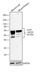
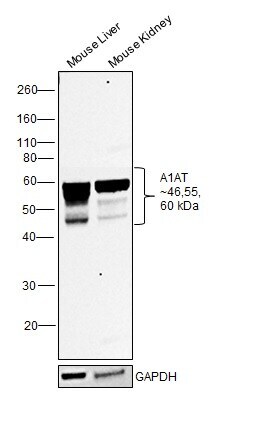
- Western blot was performed using Anti-alpha-1 Antitrypsin Monoclonal Antibody (TMF1#4B5) (Product # MIA1101) and 46, 55 and 60 kDa bands corresponding to alpha-1 Antitrypsin and its glycosylated forms were observed across tissues tested. Membrane enriched extracts (30 µg lysate) of Mouse Liver (Lane 1) and Mouse Kidney (Lane 2) were electrophoresed using NuPAGE™ 10% Bis-Tris Protein Gel (Product # NP0302BOX). Resolved proteins were then transferred onto a Nitrocellulose membrane (Product # IB23001) by iBlot® 2 Dry Blotting System (Product # IB21001). The blot was probed with the primary antibody (2 µg/mL) and detected by chemiluminescence with Goat anti-Mouse IgG (H+L) Superclonal™ Recombinant Secondary Antibody, HRP (Product # A28177, 1:4000 dilution) using the iBright FL 1000 (Product # A32752). Chemiluminescent detection was performed using Novex® ECL Chemiluminescent Substrate Reagent Kit (Product # WP20005).
Supportive validation
- Submitted by
- Invitrogen Antibodies (provider)
- Main image

- Experimental details
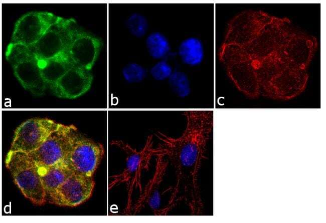
- Immunofluorescent analysis of Alpha1-Antitrypsin was performed using 70% confluent log phase Hep G2 cells. The cells were fixed with 4% paraformaldehyde for 10 minutes, permeabilized with 0.1% Triton™ X-100 for 10 minutes, and blocked with 1% BSA for 1 hour at room temperature. The cells were labeled with Alpha1-Antitrypsin (TMF1#4B5) Mouse Monoclonal Antibody (Product # MIA1101) at 2 µg/mL in 0.1% BSA and incubated for 3 hours at room temperature and then labeled with Goat anti-Mouse IgG (H+L) Superclonal™ Secondary Antibody, Alexa Fluor® 488 conjugate (Product # A28175) a dilution of 1:2000 for 45 minutes at room temperature (Panel a: green). Nuclei (Panel b: blue) were stained with SlowFade® Gold Antifade Mountant with DAPI (Product # S36938). F-actin (Panel c: red) was stained with Alexa Fluor® 555 Rhodamine Phalloidin (Product # R415, 1:300). Panel d represents the merged image showing cytoplasmic localization. Panel e shows the no primary antibody control. The images were captured at 60X magnification.