 Explore
 Validate
 Learn
Learn Western blot
Antibody data
- Antibody Data
- Antigen structure
- References [0]
- Comments [0]
- Validations
- Western blot [4]
- Immunocytochemistry [2]
- Immunohistochemistry [1]
Submit
Validation data
Reference
Comment
Report error
- Product number
- PA5-60717 - Provider product page

- Provider
- Invitrogen Antibodies
- Product name
- SHARPIN Polyclonal Antibody
- Antibody type
- Polyclonal
- Antigen
- Recombinant full-length protein
- Description
- Immunogen sequence: KSNSPPALGP EACPVSLPSP PEASTLKGPP PEADLPRSPG NLTEREELAG SLARAIAGGD EKGAAQVAAV LAQHRVALSV QLQEACFPPG
- Concentration
- 0.2 mg/mL
No comments: Submit comment
Supportive validation
- Submitted by
- Invitrogen Antibodies (provider)
- Main image
- Experimental details
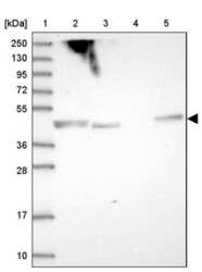
- Western blot analysis of SHARPIN in Lane 1: Marker (kDa) 250, 130, 95, 72, 55, 36, 28, 17, 10; Lane 2: Human cell line RT-4; Lane 3: Human cell line U-251MG sp; Lane 4: Human plasma (IgG/HSA depleted); Lane 5: Human liver tissue. Samples were probed using a SHARPIN Polyclonal Antibody (Product # PA5-60717).

- Submitted by
- Invitrogen Antibodies (provider)
- Main image
- Experimental details
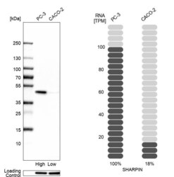
- Western blot analysis of SHARPin in human cell lines PC-3 and Caco-2 using a SHARPIN Polyclonal Antibody (Product # PA5-60717). Corresponding SHARPIN RNA-seq data are presented for the same cell lines. Loading control: Anti-HSP90B1.

- Submitted by
- Invitrogen Antibodies (provider)
- Main image

- Experimental details
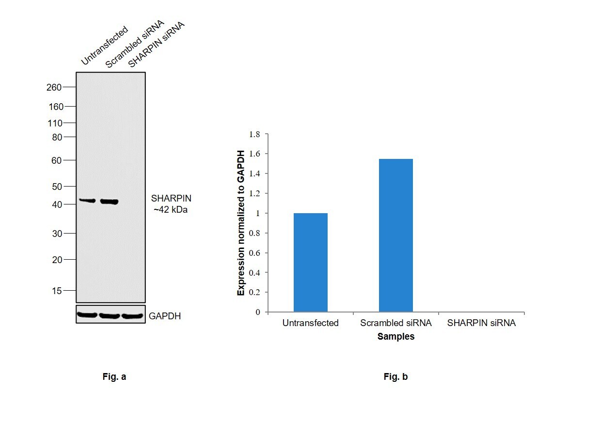
- Knockdown of SHARPIN was achieved by transfecting A549 with SHARPIN specific siRNAs (Silencer® select Product # s195427, s37848). Western blot analysis (Fig. a) was performed using Whole cell extracts from the SHARPIN knockdown cells (lane 3), non-targeting scrambled siRNA transfected cells (lane 2) and untransfected cells (lane 1). The blot was probed with SHARPIN Polyclonal Antibody (Product # PA5-60717, 0.1 µg/mL) and Goat anti-Rabbit IgG (H+L) Superclonal™ Recombinant Secondary Antibody, HRP (Product # A27036, 1:4000 dilution). Densitometric analysis of this western blot is shown in histogram (Fig. b). Decrease in signal upon siRNA mediated knock down confirms that antibody is specific to SHARPIN.
- Submitted by
- Invitrogen Antibodies (provider)
- Main image
- Experimental details
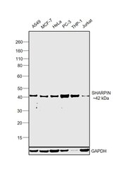
- Western blot was performed using Anti-SHARPIN Polyclonal Antibody (Product # PA5-60717) and a 42 kDa band corresponding to SHARPIN was observed across cell lines tested. Whole cell extracts (30 µg lysate) of A549 (Lane 1), MCF7 (Lane 2), HeLa (Lane 3), PC-3 (Lane 4), THP-1 (Lane 5) and Jurkat (Lane 6) were electrophoresed using NuPAGE™ 4-12% Bis-Tris Protein Gel (Product # NP0322BOX). Resolved proteins were then transferred onto a Nitrocellulose membrane (Product # IB23001) by iBlot® 2 Dry Blotting System (Product # IB21001). The blot was probed with the primary antibody (0.1 µg/mL) and detected by chemiluminescence with Goat anti-Rabbit IgG (H+L) Superclonal™ Recombinant Secondary Antibody, HRP (Product # A27036, 1:4000 dilution) using the iBright FL 1000 (Product # A32752). Chemiluminescent detection was performed using Novex® ECL Chemiluminescent Substrate Reagent Kit (Product # WP20005).

Supportive validation
- Submitted by
- Invitrogen Antibodies (provider)
- Main image
- Experimental details
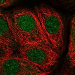
- Immunofluorescent staining of SHARPIN in human cell line MCF7 shows positivity in nucleus & cytoplasm. Samples were probed using a SHARPIN Polyclonal Antibody (Product # PA5-60717).

- Submitted by
- Invitrogen Antibodies (provider)
- Main image

- Experimental details
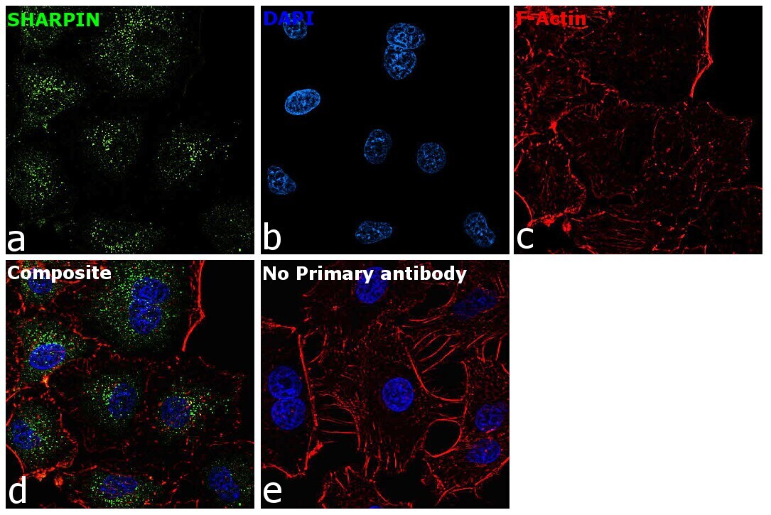
- Immunofluorescence analysis of SHARPIN was performed using 70% confluent log phase A549 cells. The cells were fixed with 4% paraformaldehyde for 10 minutes, permeabilized with 0.1% Triton™ X-100 for 15 minutes, and blocked with 2% BSA for 45 minutes at room temperature. The cells were labeled with SHARPIN Polyclonal Antibody (Product # PA5-60717) at 5 µg/mL in 0.1% BSA, incubated at 4 degree celsius overnight and then labeled with Donkey anti-Rabbit IgG (H+L) Highly Cross-Adsorbed Secondary Antibody, Alexa Fluor Plus 488 (Product # A32790), (1:2000), for 45 minutes at room temperature (Panel a: Green). Nuclei (Panel b:Blue) were stained with ProLong™ Diamond Antifade Mountant with DAPI (Product # P36962). F-actin (Panel c: Red) was stained with Rhodamine Phalloidin (Product # R415, 1:300). Panel d represents the merged image showing cytoplasmic(endosomal like pattern) localization. Panel e represents control cells with no primary antibody to assess background. The images were captured at 60X magnification.
Supportive validation
- Submitted by
- Invitrogen Antibodies (provider)
- Main image
- Experimental details
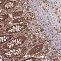
- Immunohistochemical staining of SHARPIN in human colon tissue shows strong cytoplasmic positivity in glandular cells. Samples were probed using a SHARPIN Polyclonal Antibody (Product # PA5-60717).
