Explore
 Validate
 Learn
Learn Western blot
 Immunocytochemistry
Antibody data
- Antibody Data
- Antigen structure
- References [1]
- Comments [0]
- Validations
- Western blot [1]
- Blocking/Neutralizing [1]
Submit
Validation data
Reference
Comment
Report error
- Product number
- AB-201-NA - Provider product page

- Provider
- Novus Biologicals
- Product name
- Goat Polyclonal IL-1 beta/IL-1F2 Antibody
- Antibody type
- Polyclonal
- Description
- Protein A or G purified. Detects human IL-1 beta/IL-1F2 in ELISAs and Western blots. In direct ELISAs, 100% cross-reactivity with recombinant rhesus monkey is observed, and approximately 50% cross-reactivity with recombinant mouse IL-1 beta , recombinant rat IL-1 beta , recombinant canine IL-1 beta , recombinant equine IL-1 beta , recombinant feline IL-1 beta , and approximately 20% cross-reactivity with recombinant porcine IL-1 beta and recombinant cotton rat IL-1 beta is observed.
- Reactivity
- Human
- Host
- Goat
- Conjugate
- Unconjugated
- Isotype
- IgG
- Vial size
- 1 mg
- Concentration
- LYOPH
- Storage
- Use a manual defrost freezer and avoid repeated freeze-thaw cycles. 12 months from date of receipt, -20 to -70 degreesC as supplied. 1 month, 2 to 8 degreesC under sterile conditions after reconstitution. 6 months, -20 to -70 degreesC under sterile conditions after reconstitution.
Submitted references CD40-ligand stimulates myelopoiesis by regulating flt3-ligand and thrombopoietin production in bone marrow stromal cells.
Solanilla A, Déchanet J, El Andaloussi A, Dupouy M, Godard F, Chabrol J, Charbord P, Reiffers J, Nurden AT, Weksler B, Moreau JF, Ripoche J
Blood 2000 Jun 15;95(12):3758-64
Blood 2000 Jun 15;95(12):3758-64
No comments: Submit comment
Supportive validation
- Submitted by
- Novus Biologicals (provider)
- Main image
- Experimental details
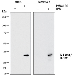
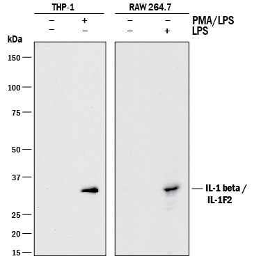
- Detection of Human and Mouse IL-1 beta/IL-1F2 by Western Blot. Western blot shows lysates of THP-1 human acute monocytic leukemia cell line untreated (-) or treated (+) with PMA and LPS and RAW 264.7 mouse monocyte/macrophage cell line untreated (-) or treated (+) with LPS. PVDF membrane was probed with 1 µg/mL of Goat Anti-Human IL-1 beta/IL-1F2 Polyclonal Antibody (Catalog # AB-201-NA) followed by HRP-conjugated Anti-Goat IgG Secondary Antibody (Catalog # HAF017). A specific band was detected for IL-1 beta/IL-1F2 at approximately 35 kDa (as indicated). This experiment was conducted under reducing conditions and using Immunoblot Buffer Group 1.
Supportive validation
- Submitted by
- Novus Biologicals (provider)
- Main image
- Experimental details
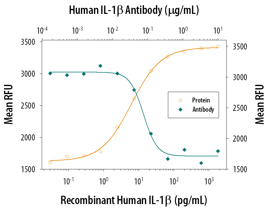
- Cell Proliferation Induced by IL-1 beta/IL-1F2 and Neutral-ization by Human IL-1 beta/ IL-1F2 Antibody. Recombinant Human IL-1 beta/IL-1F2 (Catalog # 201-LB) stimulates proliferation in the the D10.G4.1 mouse helper T cell line in a dose-dependent manner (orange line). Proliferation elicited by Recombinant Human IL-1 beta/ IL-1F2 (50 pg/mL) is neutral-ized (green line) by increasing concentrations of Goat Anti-Human IL-1 beta/IL-1F2 Poly-clonal Antibody (Catalog # AB-201-NA). The ND50 is typically 0.05-0.1 µg/mL in the presence of concanavalin A (1.25 µg/mL).