 Explore
 Validate
 Learn
Learn Western blot
Antibody data
- Antibody Data
- Antigen structure
- References [6]
- Comments [0]
- Validations
- Western blot [1]
- Immunohistochemistry [1]
Submit
Validation data
Reference
Comment
Report error
- Product number
- 14-9779-80 - Provider product page

- Provider
- Invitrogen Antibodies
- Product name
- Arginase 1 Monoclonal Antibody (sl6arg), eBioscience™
- Antibody type
- Monoclonal
- Antigen
- Other
- Description
- Immunogen sequence: MFNVMVDAK AQSTKLCSME MGQEFAKMWH QYHSKIDELI EETVKEMITL LVAKFVTILE GVLAKLSRYD EGTLFSSFLS FTVKAASKYV DVPKPGMDVA DAYVTFVRHS QDVLRDKVNE EMYIERLFDQ WYNSSMNVIC TWLTDRMDLQ LHIYQLKTLI RMVKKTYRDF RLQGVLDSTL NSKTYETIRN RLTVEEATAS VSEGGGLQGI SMKDSDEEDE EDD (1-222 aa encoded by BC015754)
- Reactivity
- Human
- Host
- Mouse
- Isotype
- IgG
- Antibody clone number
- sl6arg
- Vial size
- 25 µg
- Concentration
- 0.5 mg/mL
- Storage
- 4° C
Submitted references Deficient Adipogenesis of Scleroderma Patient and Healthy African American Monocytes.
Metabolism of L-arginine by myeloid-derived suppressor cells in cancer: mechanisms of T cell suppression and therapeutic perspectives.
Arginase-1 is a more sensitive marker of hepatic differentiation than HepPar-1 and glypican-3 in fine-needle aspiration biopsies.
Characterization of cytokine-induced myeloid-derived suppressor cells from normal human peripheral blood mononuclear cells.
Arginase-1-expressing macrophages suppress Th2 cytokine-driven inflammation and fibrosis.
Alternative metabolic states in murine macrophages reflected by the nitric oxide synthase/arginase balance: competitive regulation by CD4+ T cells correlates with Th1/Th2 phenotype.
Lee R, Reese C, Carmen-Lopez G, Perry B, Bonner M, Zemskova M, Wilson CL, Helke KL, Silver RM, Hoffman S, Tourkina E
Frontiers in pharmacology 2017;8:174
Frontiers in pharmacology 2017;8:174
Metabolism of L-arginine by myeloid-derived suppressor cells in cancer: mechanisms of T cell suppression and therapeutic perspectives.
Raber P, Ochoa AC, Rodríguez PC
Immunological investigations 2012;41(6-7):614-34
Immunological investigations 2012;41(6-7):614-34
Arginase-1 is a more sensitive marker of hepatic differentiation than HepPar-1 and glypican-3 in fine-needle aspiration biopsies.
Fujiwara M, Kwok S, Yano H, Pai RK
Cancer cytopathology 2012 Aug 25;120(4):230-7
Cancer cytopathology 2012 Aug 25;120(4):230-7
Characterization of cytokine-induced myeloid-derived suppressor cells from normal human peripheral blood mononuclear cells.
Lechner MG, Liebertz DJ, Epstein AL
Journal of immunology (Baltimore, Md. : 1950) 2010 Aug 15;185(4):2273-84
Journal of immunology (Baltimore, Md. : 1950) 2010 Aug 15;185(4):2273-84
Arginase-1-expressing macrophages suppress Th2 cytokine-driven inflammation and fibrosis.
Pesce JT, Ramalingam TR, Mentink-Kane MM, Wilson MS, El Kasmi KC, Smith AM, Thompson RW, Cheever AW, Murray PJ, Wynn TA
PLoS pathogens 2009 Apr;5(4):e1000371
PLoS pathogens 2009 Apr;5(4):e1000371
Alternative metabolic states in murine macrophages reflected by the nitric oxide synthase/arginase balance: competitive regulation by CD4+ T cells correlates with Th1/Th2 phenotype.
Munder M, Eichmann K, Modolell M
Journal of immunology (Baltimore, Md. : 1950) 1998 Jun 1;160(11):5347-54
Journal of immunology (Baltimore, Md. : 1950) 1998 Jun 1;160(11):5347-54
No comments: Submit comment
Supportive validation
- Submitted by
- Invitrogen Antibodies (provider)
- Main image

- Experimental details
- Western blot was performed using Anti-Arginase 1 Monoclonal Antibody (sl6arg), eBioscience™ (Product # 14-9779-82) and a 35kDa band corresponding to Arginase-1 was observed. Whole cell extracts (40 µg lysate) of MOLT-4 (Lane 1) and Hep G2 (Lane 2) were electrophoresed using NuPAGE™ 4-12% Bis-Tris Protein Gel (Product # NP0322BOX). Resolved proteins were then transferred onto a Nitrocellulose membrane (Product # IB23001) by iBlot® 2 Dry Blotting System (Product # IB21001). The blot was probed with the primary antibody (1:500) and detected by chemiluminescence with Goat anti-Mouse IgG (H+L) Superclonal™ Recombinant Secondary Antibody, HRP (Product # A28177, 1:4000) using the iBright FL 1000 (Product # A32752). Chemiluminescent detection was performed using SuperSignal™ West Dura Extended Duration Substrate (Product # 34076).

Supportive validation
- Submitted by
- Invitrogen Antibodies (provider)
- Main image
- Experimental details
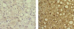
- Immunohistochemistry of formalin-fixed paraffin embedded human liver tissue using 5 µg/mL of Mouse IgG1 K Isotype Control Purified (Product # 14-4714-82) (left) or 5 µg/mL of Anti-Human Arginase-1 Purified (right), followed by Anti-Mouse IgG Biotin, Streptavidin HRP, and DAB visualization.Nuclei are counterstained with hematoxylin.
