 Explore
 Validate
 Learn
Learn Western blot
 ELISA
ELISAAntibody data
- Antibody Data
- Antigen structure
- References [9]
- Comments [0]
- Validations
- Western blot [4]
- Other assay [16]
Submit
Validation data
Reference
Comment
Report error
- Product number
- 33-1520 - Provider product page

- Provider
- Invitrogen Antibodies
- Product name
- Occludin Monoclonal Antibody (OC-3F10), HRP
- Antibody type
- Monoclonal
- Antigen
- Other
- Reactivity
- Human, Mouse, Rat, Canine
- Host
- Mouse
- Conjugate
- Horseradish Peroxidase
- Isotype
- IgG
- Antibody clone number
- OC-3F10
- Vial size
- 100 µg
- Concentration
- 1 mg/mL
- Storage
- 4° C
Submitted references Astrocyte-derived VEGF increases cerebral microvascular permeability under high salt conditions.
Fingolimod reduces circulating tight-junction protein levels and in vitro peripheral blood mononuclear cells migration in multiple sclerosis patients.
TNFα alters occludin and cerebral endothelial permeability: Role of p38MAPK.
12/15-Lipoxygenase-dependent ROS production is required for diet-induced endothelial barrier dysfunction.
HIV-1 Tat C modulates expression of miRNA-101 to suppress VE-cadherin in human brain microvascular endothelial cells.
Differential effects of flavonoids on barrier integrity in human intestinal Caco-2 cells.
Protein kinase Cζ phosphorylates occludin and promotes assembly of epithelial tight junctions.
Role of megalin (gp330) in transcytosis of thyroglobulin by thyroid cells. A novel function in the control of thyroid hormone release.
Role of megalin (gp330) in transcytosis of thyroglobulin by thyroid cells. A novel function in the control of thyroid hormone release.
Deng Z, Zhou L, Wang Y, Liao S, Huang Y, Shan Y, Tan S, Zeng Q, Peng L, Huang H, Lu Z
Aging 2020 Jun 22;12(12):11781-11793
Aging 2020 Jun 22;12(12):11781-11793
Fingolimod reduces circulating tight-junction protein levels and in vitro peripheral blood mononuclear cells migration in multiple sclerosis patients.
Annunziata P, Cioni C, Masi G, Tassi M, Marotta G, Severi S
Scientific reports 2018 Oct 18;8(1):15371
Scientific reports 2018 Oct 18;8(1):15371
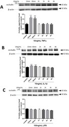
TNFα alters occludin and cerebral endothelial permeability: Role of p38MAPK.
Ni Y, Teng T, Li R, Simonyi A, Sun GY, Lee JC
PloS one 2017;12(2):e0170346
PloS one 2017;12(2):e0170346
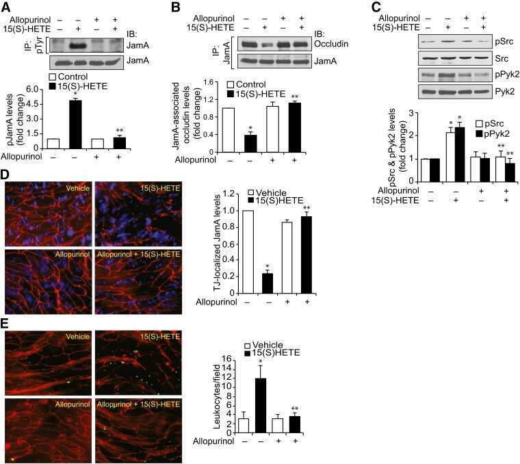
12/15-Lipoxygenase-dependent ROS production is required for diet-induced endothelial barrier dysfunction.
Chattopadhyay R, Tinnikov A, Dyukova E, Singh NK, Kotla S, Mobley JA, Rao GN
Journal of lipid research 2015 Mar;56(3):562-577
Journal of lipid research 2015 Mar;56(3):562-577
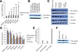
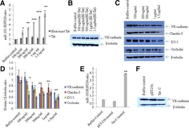
HIV-1 Tat C modulates expression of miRNA-101 to suppress VE-cadherin in human brain microvascular endothelial cells.
Mishra R, Singh SK
The Journal of neuroscience : the official journal of the Society for Neuroscience 2013 Apr 3;33(14):5992-6000
The Journal of neuroscience : the official journal of the Society for Neuroscience 2013 Apr 3;33(14):5992-6000
Differential effects of flavonoids on barrier integrity in human intestinal Caco-2 cells.
Noda S, Tanabe S, Suzuki T
Journal of agricultural and food chemistry 2012 May 9;60(18):4628-33
Journal of agricultural and food chemistry 2012 May 9;60(18):4628-33
Protein kinase Cζ phosphorylates occludin and promotes assembly of epithelial tight junctions.
Jain S, Suzuki T, Seth A, Samak G, Rao R
The Biochemical journal 2011 Jul 15;437(2):289-99
The Biochemical journal 2011 Jul 15;437(2):289-99
Role of megalin (gp330) in transcytosis of thyroglobulin by thyroid cells. A novel function in the control of thyroid hormone release.
Marinò M, Zheng G, Chiovato L, Pinchera A, Brown D, Andrews D, McCluskey RT
The Journal of biological chemistry 2000 Mar 10;275(10):7125-37
The Journal of biological chemistry 2000 Mar 10;275(10):7125-37
No comments: Submit comment
Supportive validation
- Submitted by
- Invitrogen Antibodies (provider)
- Main image
- Experimental details
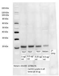
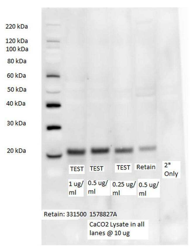
- Western blot analysis of Occludin was performed by loading 10 µg of CaCO2 lysate per well onto a 4-12% Bis-Tris polyacrylamide gel. Proteins were transferred to a nitrocellulose membrane and blocked with 5% BSA/TBST for at least 1 hour. The membrane was probed with an Occludin purified monoclonal antibody (Product # 33-1500) at a dilution of 1 µg/mL, 0.5 µg/mL, and 0.25 µg/mL for 1 hour at room temperature on a rocking platform and washed in TBS-0.1% Tween-20. Goat Anti-Mouse IGG (H+L) HRP Conjugated secondary antibody (Product # 31430) was used at a concentration of 1:20,000 and incubated for 30 minutes on a rocking platform. Super Signal West Dura substrate (Product # 34076) was incubated for 5 minutes on a rocking platform. Detection was performed using the GBox. The 5th isoform of Occludin was detected at ~23 kDa.
- Conjugate
- Horseradish Peroxidase
- Submitted by
- Invitrogen Antibodies (provider)
- Main image

- Experimental details
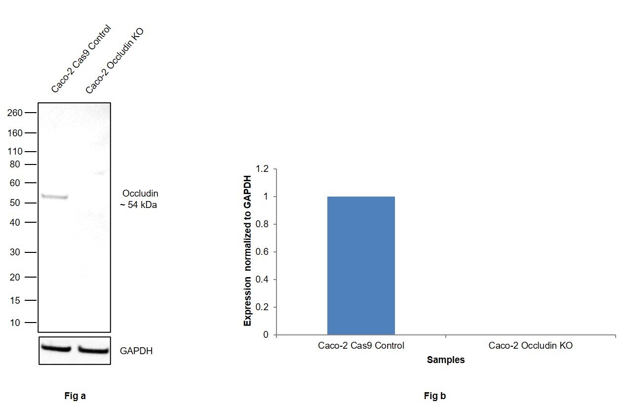
- Knockout of Occludin was achieved by CRISPR-Cas9 genome editing using LentiArray™ Lentiviral sgRNA (Product # A32042, Assay ID CRISPR777079_LV) and LentiArray Cas9 Lentivirus (Product # A32064). Western blot analysis of Occludin was performed by loading 30 µg of Caco-2 Cas9 (Lane 1) andCaco-2 Occludin KO (Lane 2) membrane enriched extracts. The samples were electrophoresed using NuPAGE™ Novex™ 4-12% Bis-Tris Protein Gel (Product # NP0322BOX). Resolved proteins were then transferred onto a nitrocellulose membrane (Product # IB23001) by iBlot® 2 Dry Blotting System (Product # IB21001). The blot was probed with Anti-Occludin Monoclonal Antibody-HRP (OC-3F10) (Product # 33-1520, 1:500 dilution) using the iBright FL 1000 (Product # A32752). Chemiluminescent detection was performed using Novex® ECL Chemiluminescent Substrate Reagent Kit (Product # WP20005). Loss of signal upon CRISPR mediated knockout (KO) using the LentiArray™ CRISPR product line confirms that antibody is specific to Occludin.
- Conjugate
- Horseradish Peroxidase
- Submitted by
- Invitrogen Antibodies (provider)
- Main image

- Experimental details
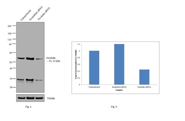
- Knockdown of Occludin was achieved by transfecting Caco-2 with Occludin specific siRNAs (Silencer® select Product # s9812, s9814). Western blot analysis (Fig. a) was performed using whole cell extracts from the Occludin knockdown cells (lane 3), non-specific scrambled siRNA transfected cells (lane 2) and untransfected cells (lane 1). The blots were probed with Occludin Monoclonal Antibody (OC-3F10), HRP (Product # 33-1520, 2µg/ml). Densitometric analysis of this western blot is shown in histogram (Fig. b). Decrease in signal upon siRNA mediated knock down confirms that antibody is specific to Occludin.
- Conjugate
- Horseradish Peroxidase
- Submitted by
- Invitrogen Antibodies (provider)
- Main image

- Experimental details
- Western blot analysis was performed on whole cell extracts (30 µg lysate) of Caco-2 (Lane 1) and Caco-2 treated with IL 1 beta (10 ng/ml for 48 hr) (Lane 2). The blot was probed with Anti-Occludin Monoclonal Antibody (OC-3F10), HRP (Product # 33-1520, 2 µg/ml). A 54, 30 kDa bands corresponding to Occludin was observed in the cell line tested and decreased upon treatment.
- Conjugate
- Horseradish Peroxidase

Supportive validation
- Submitted by
- Invitrogen Antibodies (provider)
- Main image

- Experimental details
- NULL
- Conjugate
- Horseradish Peroxidase
- Submitted by
- Invitrogen Antibodies (provider)
- Main image

- Experimental details
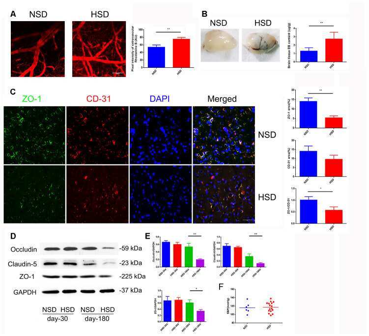
- Figure 1 HSD increases cerebral microvascular permeability. ( A ) Representative images of the mice cerebral cortical microvessels and dye leakage at 10 min after injection of Rhodamine B isothiocyanate-dextran (n=3 per group). ( B ) Evans blue leakage analyzed in rats fed with selected diets for 180 days (n=3 per group). ( C ) ZO-1 (green) co-stained with CD31 (red), a microvascular endothelia marker, and DAPI (blue; n=6 per group) in the rats brain slices. ( D , E ) Expression of Occludin, Claudin-5 and ZO-1 in the brain tissues from rats analyzed by immunoblotting. ( F ) Systolic blood pressure (SBP) of rats fed with NSD or HSD for 180 days. * P
- Conjugate
- Horseradish Peroxidase
- Submitted by
- Invitrogen Antibodies (provider)
- Main image

- Experimental details
- NULL
- Conjugate
- Horseradish Peroxidase
- Submitted by
- Invitrogen Antibodies (provider)
- Main image
- Experimental details
- NULL
- Conjugate
- Horseradish Peroxidase
- Submitted by
- Invitrogen Antibodies (provider)
- Main image
- Experimental details
- NULL
- Conjugate
- Horseradish Peroxidase
- Submitted by
- Invitrogen Antibodies (provider)
- Main image
- Experimental details
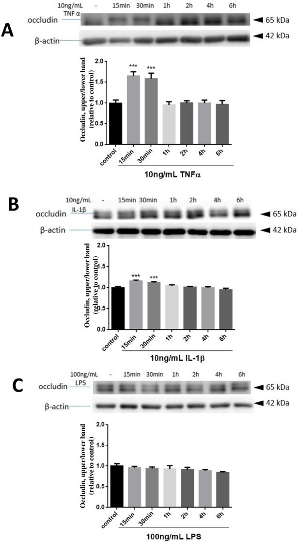
- Fig 2 Effects of TNFalpha, IL-1beta and LPS on occludin band-shift in hCMEC/D3 cells. Cells were treated with or without TNFalpha (10 ng/mL) (A), IL-1beta (10 ng/mL)(B) and LPS (100 ng/mL) (C) for 15, 30 min and 1, 2, 4, 6 hours. Cell lysates were collected and occludin and beta-actin expression patterns were analyzed by immunoblotting assay as described in text. The density of upper band versus total upper and lower and normalized with beta-actin was determined by measuring protein intensity as PIupper/PItotal/PIbeta-actin. Results are mean +- SD from 4 or more experiments and data are analyzed by one-way ANOVA followed by Bonferroni post-tests (***P
- Conjugate
- Horseradish Peroxidase
- Submitted by
- Invitrogen Antibodies (provider)
- Main image

- Experimental details
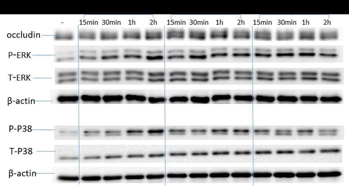
- Fig 4 TNFalpha-mediated occludin band-shift and phosphorylation of ERK1/2 and p38MAPK in hCMEC/D3 cells. Cells were treated with or without TNFalpha (10 ng/mL) at 5, 10, 15, 30, 60 min (A). Cell lysates were collected and occludin, P- ERK, T-ERK, P-P38, T-P38 and beta-actin expression pattern was analyzed by immunoblotting assay. Quantification of the proportion of occludin upper band were determined through PI upper /PI total /PI beta-actin and then normalized to control (B). phospho-ERK1/2 (C) and phospho-p38MAPK (D) were similarly quantified and plotted. Results are mean +- SD from 3 or more experiments and data are analyzed by one-way ANOVA followed by Bonferroni post-tests (**P
- Conjugate
- Horseradish Peroxidase

- Submitted by
- Invitrogen Antibodies (provider)
- Main image
- Experimental details
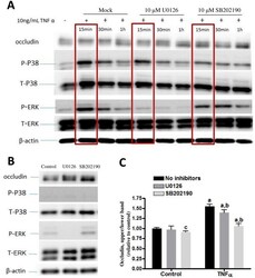
- Fig 5 Effects of p-ERK1/2 and p-p38MAPK inhibitors on occludin band-shift in CMEC/D3 cells. Cells were pretreated with or without 10 muM U0126 and 10 muM SB202190 for 15 min and then stimulated with or without 10 ng/mL TNFalpha for 15, 30, 60 min (A and C). (B) Testing the effects of inhibitors alone. Cell lysates were collected and occludin, P- ERK, T-ERK, P-p38, T-p38 and beta-actin expression pattern was analyzed by immunoblotting assay. Quantification of the proportions of occludin upper band at 15 min of TNFalpha and with inhibitors (blots enclosed in brackets) were determined through PI upper /PI total /PI beta-actin and then normalized to control. Results are mean +- SD from 4 or more experiments. Two-way ANOVA revealed significant main effects of TNFalpha and the inhibitors and a significant interaction (p
- Conjugate
- Horseradish Peroxidase

- Submitted by
- Invitrogen Antibodies (provider)
- Main image
- Experimental details
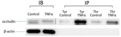
- Fig 6 Effects of TNFalpha on tyrosine and threonine phosphorylation of occludin. hCMEC/D3 cells were treated with or without 10 ng/mL TNFalpha for 15 min. Cell lysates were mixed with anti-tyrosine or anti-threonine antibody conjugated with protein A beads and then phosphorylated proteins in cell lysates were pulled down. Occludin and beta-actin expression pattern was analyzed by immunoblotting assay.
- Conjugate
- Horseradish Peroxidase

- Submitted by
- Invitrogen Antibodies (provider)
- Main image

- Experimental details
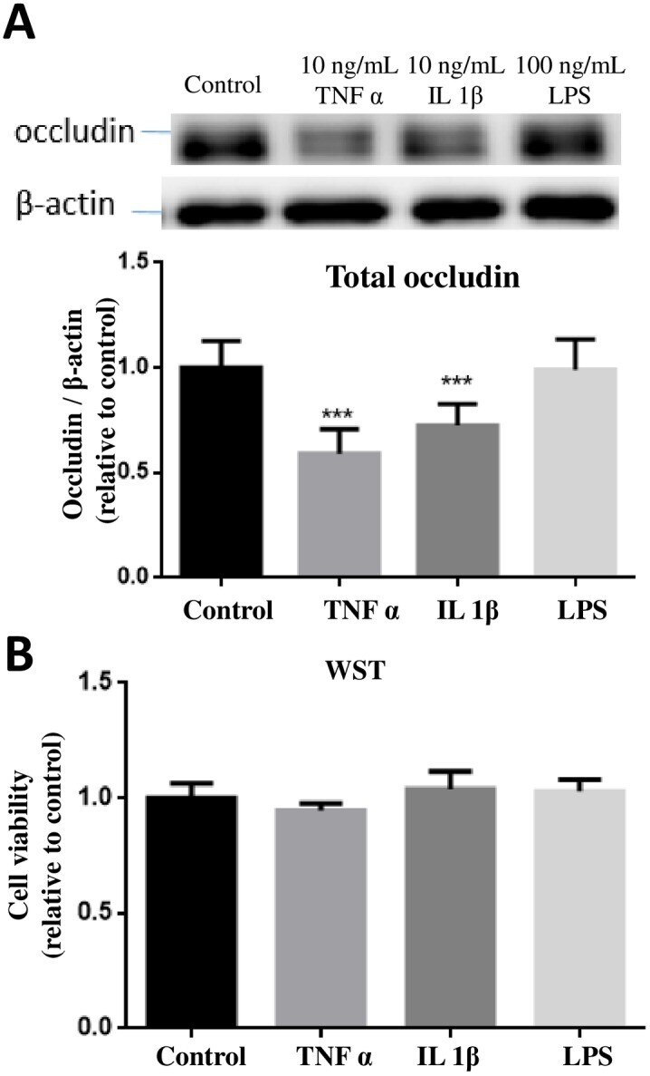
- Fig 7 Effects of TNFalpha, IL-1beta and LPS on occludin expression and cell viability. (A) hCMEC/D3 cells were treated with or without TNFalpha (10 ng/mL), IL-1beta (10 ng/mL) and LPS (100 ng/mL) for 24 hours. Cell lysates were collected and occludin and beta-actin expression pattern was analyzed by immunoblotting assay. Quantification of the protein band intensity was determined through PI total /PI beta-actin and then normalized to control. Results are mean +- SD from 4 or more experiments and data are analyzed by one-way ANOVA followed by Bonferroni post-tests (***P
- Conjugate
- Horseradish Peroxidase
- Submitted by
- Invitrogen Antibodies (provider)
- Main image

- Experimental details
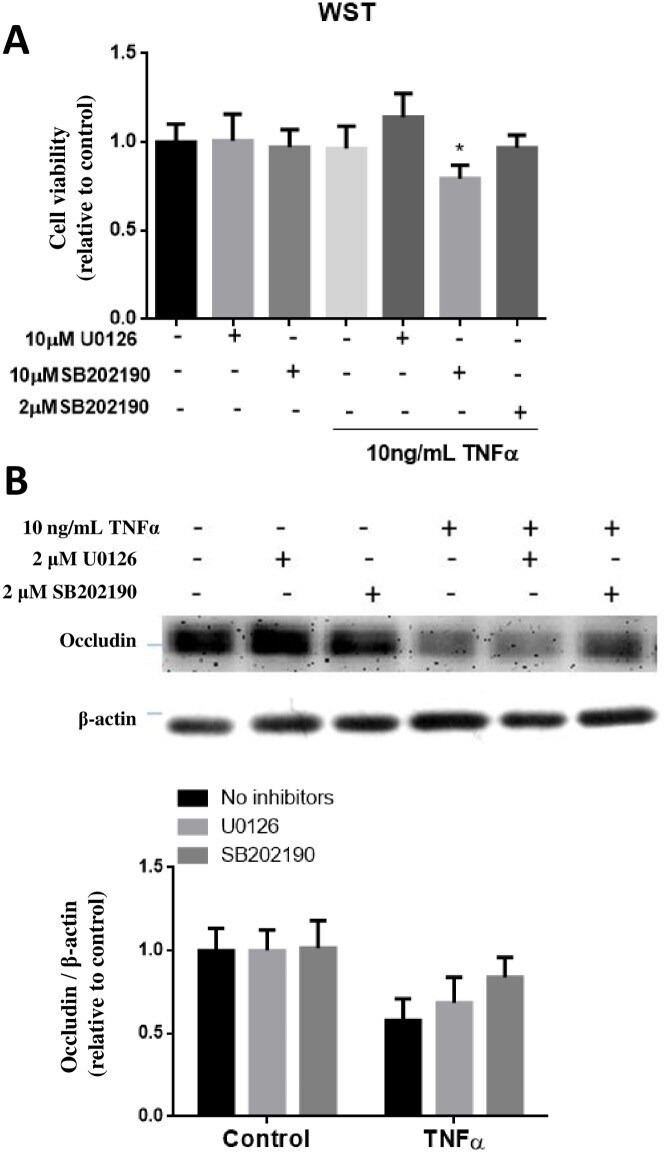
- Fig 8 Effects of p-ERK1/2 and p-p38MAPK inhibitors on TNFalpha-induced occludin expression in hCMEC/D3 cells. (A) Initial testing for cell viability using WST-1 assay indicated toxicity of cells upon incubation (24h) with TNFalpha (10 ng/mL) in the presence of SB202190 at 10 muM but not at 2 muM. (B) Testing ability of U0126 (2 muM) and SB202190 (2 muM) to ameliorate the decrease in occludin expression upon exposure of TNFalpha for 24 h. Cell lysates were collected and occludin and beta-actin expression pattern was analyzed by immunoblotting assay. Quantification of protein band intensity was determined through PItotal/PIbeta-actin and then normalized to control. Results are mean +- SD from 5 or more experiments. Two-way ANOVA revealed a significant main effect of TNFalpha (p
- Conjugate
- Horseradish Peroxidase
- Submitted by
- Invitrogen Antibodies (provider)
- Main image

- Experimental details
- Figure 1 HSD increases cerebral microvascular permeability. ( A ) Representative images of the mice cerebral cortical microvessels and dye leakage at 10 min after injection of Rhodamine B isothiocyanate-dextran (n=3 per group). ( B ) Evans blue leakage analyzed in rats fed with selected diets for 180 days (n=3 per group). ( C ) ZO-1 (green) co-stained with CD31 (red), a microvascular endothelia marker, and DAPI (blue; n=6 per group) in the rats brain slices. ( D , E ) Expression of Occludin, Claudin-5 and ZO-1 in the brain tissues from rats analyzed by immunoblotting. ( F ) Systolic blood pressure (SBP) of rats fed with NSD or HSD for 180 days. * P
- Conjugate
- Horseradish Peroxidase

- Submitted by
- Invitrogen Antibodies (provider)
- Main image
- Experimental details
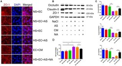
- Figure 4 Astrocyte-derived VEGF mediates HS-induced BBB breakdown. ( A ) Representative double immunofluorescence staining of ZO-1 + bEnd.3 endothelium. Endothelial cells were cultured alone, or co-cultured with primary rats' astrocytes, and treated with NS, HS, conditioned medium, or VEGF neutralizing antibody (NA). ( B , C ) Western blotting analysis of Occludin, Claudin-5, and ZO-1 in endothelial cells. ( D ) Permeability of tight junctions measured using NaF; * P
- Conjugate
- Horseradish Peroxidase

- Submitted by
- Invitrogen Antibodies (provider)
- Main image

- Experimental details
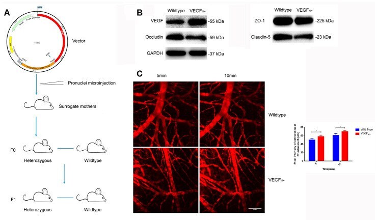
- Figure 6 Increased cerebral microvascular permeability in VEGF hi/+ mice. ( A ) Schematic diagram of VEGF hi/+ mice generation. ( B ) Immunoblotting showing VEGF, Occludin, Claudin-5 and ZO-1expression in VEGF hi/+ and wildtype mice. ( C ) Cerebral cortical micro-vessel and dye leakage at 5 and 10 min after injection of Rhodamine B isothiocyanate-dextran; n=5, * P
- Conjugate
- Horseradish Peroxidase
- Submitted by
- Invitrogen Antibodies (provider)
- Main image

- Experimental details
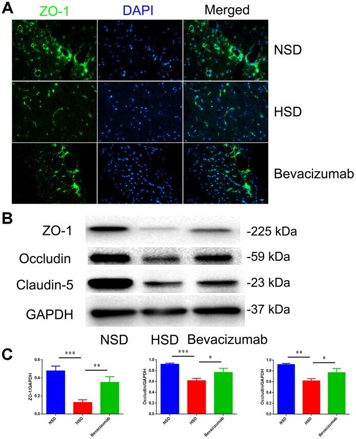
- Figure 7 Blocking VEGF attenuates disruption of tight junctions induced by HSD. ( A ) Representative images showing double immunofluorescence staining of ZO-1 in green and DAPI in the brain specimens of rats. ( B , C ) Expression of Occludin, Claudin-5 and ZO-1 determined by western blotting; n=5, * P
- Conjugate
- Horseradish Peroxidase
- Submitted by
- Invitrogen Antibodies (provider)
- Main image
- Experimental details
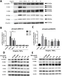
- Fig 3 Effects of TNFalpha, IL-1beta and LPS on p-ERK1/2 and p-p38MAPK expression in hCMEC/D3 cells. Cells were treated with or without TNFalpha (A, B, C), IL-1beta (D) and LPS (E) for 15, 30 min and 1, 2, 4, 6 hours. Cell lysates were collected and phospho-ERK1/2 (P- ERK), total ERK1/2 (T-ERK), phospho-p38MAPK (P-P38), total p38MAPK (T-P38) and beta-actin expression pattern were analyzed by immunoblotting assay. Quantification of phospho-proteins was determined through assay of PIphospho/PItotal/PIbeta-actin and then normalized to control. Results of (B) and (C) are mean +- SD from 4 or more experiments and data are analyzed by one-way ANOVA followed by Bonferroni post-tests **P
- Conjugate
- Horseradish Peroxidase
