 Explore
 Validate
 Learn
Learn Immunohistochemistry
Antibody data
- Antibody Data
- Antigen structure
- References [0]
- Comments [0]
- Validations
- Immunohistochemistry [35]
Submit
Validation data
Reference
Comment
Report error
- Product number
- LS-C483078 - Provider product page

- Provider
- LSBio
- Product name
- TJP2 / ZO2 / ZO-2 Antibody (aa740-790) LS-C483078
- Antibody type
- Polyclonal
- Description
- Antiserum
- Reactivity
- Human
- Host
- Rabbit
- Storage
- Maintain lyophilized and reconstituted antibodies at -20°C for long term storage and at 2°C to 8°C for a shorter term. When reconstituting, glycerol (1:1) may be added for an additional stability. Avoid freeze/thaw cycles.
No comments: Submit comment
Supportive validation
- Submitted by
- LSBio (provider)
- Enhanced method
- Genetic validation
- Main image
- Experimental details
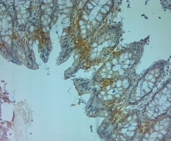
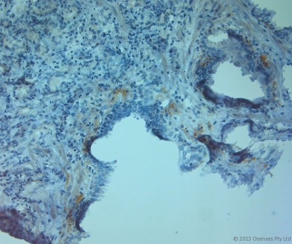
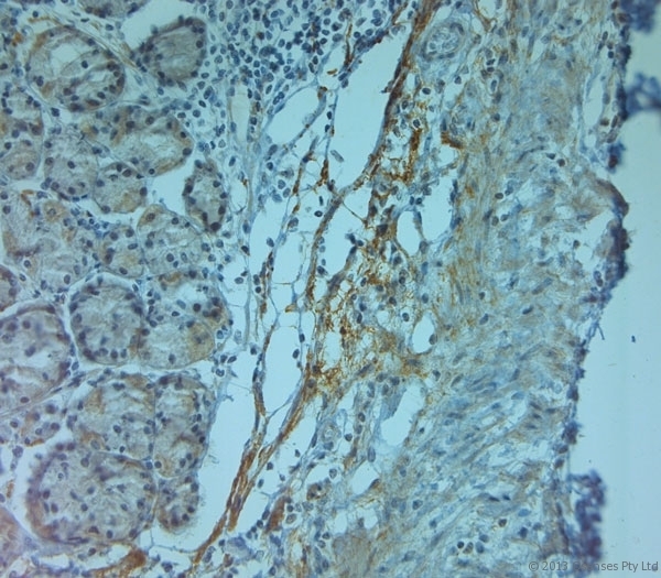
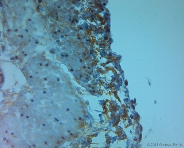
- Rabbit antibody to ZO2 (740-790). IHC on paraffin sections of human large intestine tissue using Rabbit antibody to ZO2 (740-790). HIER: 1 mM EDTA, pH 8 for 20 min using Thermo PT Module. Blocking: 0.2% LFDM in TBST filtered through a 0.2 micron filter. Detection was done using Novolink HRP polymer from Leica following manufacturer's instructions. Primary antibody: dilution 1:1000, incubated 30 min at RT (using Autostainer). Sections were counterstained with Harris Hematoxylin.

- Submitted by
- LSBio (provider)
- Enhanced method
- Genetic validation
- Main image

- Experimental details
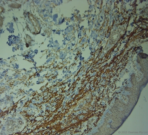
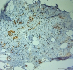
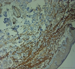
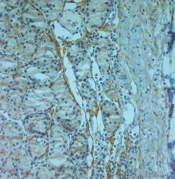
- Rabbit antibody to ZO2 (740-790). IHC on paraffin sections of human skin tissue using Rabbit antibody to ZO2 (740-790). HIER: 1 mM EDTA, pH 8 for 20 min using Thermo PT Module. Blocking: 0.2% LFDM in TBST filtered through a 0.2 micron filter. Detection was done using Novolink HRP polymer from Leica following manufacturer's instructions. Primary antibody: dilution 1:1000, incubated 30 min at RT (using Autostainer). Sections were counterstained with Harris Hematoxylin.
- Submitted by
- LSBio (provider)
- Enhanced method
- Genetic validation
- Main image

- Experimental details
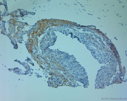
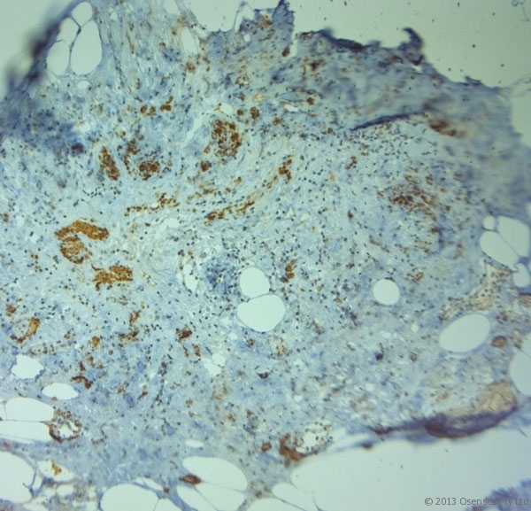
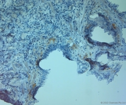
- Rabbit antibody to ZO2 (740-790). IHC on paraffin sections of human skin tissue using Rabbit antibody to ZO2 (740-790). HIER: 1 mM EDTA, pH 8 for 20 min using Thermo PT Module. Blocking: 0.2% LFDM in TBST filtered through a 0.2 micron filter. Detection was done using Novolink HRP polymer from Leica following manufacturer's instructions. Primary antibody: dilution 1:1000, incubated 30 min at RT (using Autostainer). Sections were counterstained with Harris Hematoxylin.

- Submitted by
- LSBio (provider)
- Enhanced method
- Genetic validation
- Main image
- Experimental details
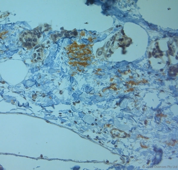
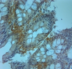
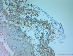
- Rabbit antibody to ZO2 (740-790). IHC on paraffin sections of human uterus tissue using Rabbit antibody to ZO2 (740-790). HIER: 1 mM EDTA, pH 8 for 20 min using Thermo PT Module. Blocking: 0.2% LFDM in TBST filtered through a 0.2 micron filter. Detection was done using Novolink HRP polymer from Leica following manufacturer's instructions. Primary antibody: dilution 1:1000, incubated 30 min at RT (using Autostainer). Sections were counterstained with Harris Hematoxylin.

- Submitted by
- LSBio (provider)
- Enhanced method
- Genetic validation
- Main image

- Experimental details
- Rabbit antibody to ZO2 (740-790). IHC on paraffin sections of human breast tissue using Rabbit antibody to ZO2 (740-790). HIER: 1 mM EDTA, pH 8 for 20 min using Thermo PT Module. Blocking: 0.2% LFDM in TBST filtered through a 0.2 micron filter. Detection was done using Novolink HRP polymer from Leica following manufacturer's instructions. Primary antibody: dilution 1:1000, incubated 30 min at RT (using Autostainer). Sections were counterstained with Harris Hematoxylin.
- Submitted by
- LSBio (provider)
- Enhanced method
- Genetic validation
- Main image

- Experimental details
- Rabbit antibody to ZO2 (740-790). IHC on paraffin sections of human prostate tissue using Rabbit antibody to ZO2 (740-790). HIER: 1 mM EDTA, pH 8 for 20 min using Thermo PT Module. Blocking: 0.2% LFDM in TBST filtered through a 0.2 micron filter. Detection was done using Novolink HRP polymer from Leica following manufacturer's instructions. Primary antibody: dilution 1:1000, incubated 30 min at RT (using Autostainer). Sections were counterstained with Harris Hematoxylin.
- Submitted by
- LSBio (provider)
- Enhanced method
- Genetic validation
- Main image

- Experimental details
- Rabbit antibody to ZO2 (740-790). IHC on paraffin sections of human stomach tissue using Rabbit antibody to ZO2 (740-790). HIER: 1 mM EDTA, pH 8 for 20 min using Thermo PT Module. Blocking: 0.2% LFDM in TBST filtered through a 0.2 micron filter. Detection was done using Novolink HRP polymer from Leica following manufacturer's instructions. Primary antibody: dilution 1:1000, incubated 30 min at RT (using Autostainer). Sections were counterstained with Harris Hematoxylin.

- Submitted by
- LSBio (provider)
- Enhanced method
- Genetic validation
- Main image

- Experimental details
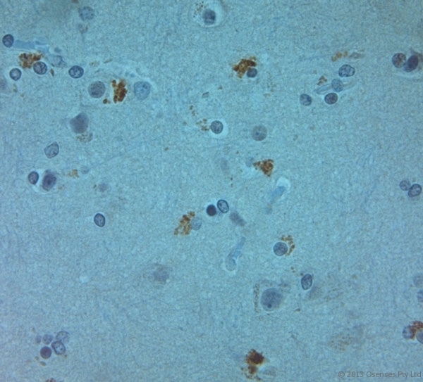
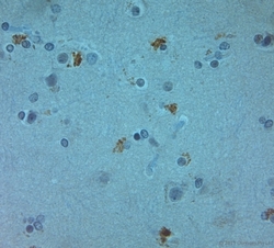
- Rabbit antibody to ZO2 (740-790). IHC on paraffin sections of human brain tissue using Rabbit antibody to ZO2 (740-790). HIER: 1 mM EDTA, pH 8 for 20 min using Thermo PT Module. Blocking: 0.2% LFDM in TBST filtered through a 0.2 micron filter. Detection was done using Novolink HRP polymer from Leica following manufacturer's instructions. Primary antibody: dilution 1:1000, incubated 30 min at RT (using Autostainer). Sections were counterstained with Harris Hematoxylin.

- Submitted by
- LSBio (provider)
- Enhanced method
- Genetic validation
- Main image
- Experimental details
- Rabbit antibody to ZO2 (740-790). IHC on paraffin sections of human appendix tissue using Rabbit antibody to ZO2 (740-790). HIER: 1 mM EDTA, pH 8 for 20 min using Thermo PT Module. Blocking: 0.2% LFDM in TBST filtered through a 0.2 micron filter. Detection was done using Novolink HRP polymer from Leica following manufacturer's instructions. Primary antibody: dilution 1:1000, incubated 30 min at RT (using Autostainer). Sections were counterstained with Harris Hematoxylin.
- Submitted by
- LSBio (provider)
- Enhanced method
- Genetic validation
- Main image

- Experimental details
- Rabbit antibody to ZO2 (740-790). IHC on paraffin sections of human large intestine tissue using Rabbit antibody to ZO2 (740-790). HIER: 1 mM EDTA, pH 8 for 20 min using Thermo PT Module. Blocking: 0.2% LFDM in TBST filtered through a 0.2 micron filter. Detection was done using Novolink HRP polymer from Leica following manufacturer's instructions. Primary antibody: dilution 1:1000, incubated 30 min at RT (using Autostainer). Sections were counterstained with Harris Hematoxylin.

- Submitted by
- LSBio (provider)
- Enhanced method
- Genetic validation
- Main image

- Experimental details
- Rabbit antibody to ZO2 (740-790). IHC on paraffin sections of human stomach tissue using Rabbit antibody to ZO2 (740-790). HIER: 1 mM EDTA, pH 8 for 20 min using Thermo PT Module. Blocking: 0.2% LFDM in TBST filtered through a 0.2 micron filter. Detection was done using Novolink HRP polymer from Leica following manufacturer's instructions. Primary antibody: dilution 1:1000, incubated 30 min at RT (using Autostainer). Sections were counterstained with Harris Hematoxylin.
- Submitted by
- LSBio (provider)
- Main image

- Experimental details
- Rabbit antibody to ZO2 (740-790). IHC on paraffin sections of human large intestine tissue using Rabbit antibody to ZO2 (740-790). HIER: 1 mM EDTA, pH 8 for 20 min using Thermo PT Module. Blocking: 0.2% LFDM in TBST filtered through a 0.2 micron filter. Detection was done using Novolink HRP polymer from Leica following manufacturer's instructions. Primary antibody: dilution 1:1000, incubated 30 min at RT (using Autostainer). Sections were counterstained with Harris Hematoxylin.

- Submitted by
- LSBio (provider)
- Main image
- Experimental details
- Rabbit antibody to ZO2 (740-790). IHC on paraffin sections of human skin tissue using Rabbit antibody to ZO2 (740-790). HIER: 1 mM EDTA, pH 8 for 20 min using Thermo PT Module. Blocking: 0.2% LFDM in TBST filtered through a 0.2 micron filter. Detection was done using Novolink HRP polymer from Leica following manufacturer's instructions. Primary antibody: dilution 1:1000, incubated 30 min at RT (using Autostainer). Sections were counterstained with Harris Hematoxylin.

- Submitted by
- LSBio (provider)
- Main image

- Experimental details
- Rabbit antibody to ZO2 (740-790). IHC on paraffin sections of human uterus tissue using Rabbit antibody to ZO2 (740-790). HIER: 1 mM EDTA, pH 8 for 20 min using Thermo PT Module. Blocking: 0.2% LFDM in TBST filtered through a 0.2 micron filter. Detection was done using Novolink HRP polymer from Leica following manufacturer's instructions. Primary antibody: dilution 1:1000, incubated 30 min at RT (using Autostainer). Sections were counterstained with Harris Hematoxylin.

- Submitted by
- LSBio (provider)
- Main image

- Experimental details
- Rabbit antibody to ZO2 (740-790). IHC on paraffin sections of human breast tissue using Rabbit antibody to ZO2 (740-790). HIER: 1 mM EDTA, pH 8 for 20 min using Thermo PT Module. Blocking: 0.2% LFDM in TBST filtered through a 0.2 micron filter. Detection was done using Novolink HRP polymer from Leica following manufacturer's instructions. Primary antibody: dilution 1:1000, incubated 30 min at RT (using Autostainer). Sections were counterstained with Harris Hematoxylin.

- Submitted by
- LSBio (provider)
- Main image
- Experimental details
- Rabbit antibody to ZO2 (740-790). IHC on paraffin sections of human prostate tissue using Rabbit antibody to ZO2 (740-790). HIER: 1 mM EDTA, pH 8 for 20 min using Thermo PT Module. Blocking: 0.2% LFDM in TBST filtered through a 0.2 micron filter. Detection was done using Novolink HRP polymer from Leica following manufacturer's instructions. Primary antibody: dilution 1:1000, incubated 30 min at RT (using Autostainer). Sections were counterstained with Harris Hematoxylin.

- Submitted by
- LSBio (provider)
- Main image
- Experimental details
- Rabbit antibody to ZO2 (740-790). IHC on paraffin sections of human stomach tissue using Rabbit antibody to ZO2 (740-790). HIER: 1 mM EDTA, pH 8 for 20 min using Thermo PT Module. Blocking: 0.2% LFDM in TBST filtered through a 0.2 micron filter. Detection was done using Novolink HRP polymer from Leica following manufacturer's instructions. Primary antibody: dilution 1:1000, incubated 30 min at RT (using Autostainer). Sections were counterstained with Harris Hematoxylin.

- Submitted by
- LSBio (provider)
- Main image

- Experimental details
- Rabbit antibody to ZO2 (740-790). IHC on paraffin sections of human stomach tissue using Rabbit antibody to ZO2 (740-790). HIER: 1 mM EDTA, pH 8 for 20 min using Thermo PT Module. Blocking: 0.2% LFDM in TBST filtered through a 0.2 micron filter. Detection was done using Novolink HRP polymer from Leica following manufacturer's instructions. Primary antibody: dilution 1:1000, incubated 30 min at RT (using Autostainer). Sections were counterstained with Harris Hematoxylin.
- Submitted by
- LSBio (provider)
- Main image

- Experimental details
- Rabbit antibody to ZO2 (740-790). IHC on paraffin sections of human brain tissue using Rabbit antibody to ZO2 (740-790). HIER: 1 mM EDTA, pH 8 for 20 min using Thermo PT Module. Blocking: 0.2% LFDM in TBST filtered through a 0.2 micron filter. Detection was done using Novolink HRP polymer from Leica following manufacturer's instructions. Primary antibody: dilution 1:1000, incubated 30 min at RT (using Autostainer). Sections were counterstained with Harris Hematoxylin.
- Submitted by
- LSBio (provider)
- Main image

- Experimental details
- Rabbit antibody to ZO2 (740-790). IHC on paraffin sections of human appendix tissue using Rabbit antibody to ZO2 (740-790). HIER: 1 mM EDTA, pH 8 for 20 min using Thermo PT Module. Blocking: 0.2% LFDM in TBST filtered through a 0.2 micron filter. Detection was done using Novolink HRP polymer from Leica following manufacturer's instructions. Primary antibody: dilution 1:1000, incubated 30 min at RT (using Autostainer). Sections were counterstained with Harris Hematoxylin.

- Submitted by
- LSBio (provider)
- Main image
- Experimental details
- Rabbit antibody to ZO2 (740-790). IHC on paraffin sections of human large intestine tissue using Rabbit antibody to ZO2 (740-790). HIER: 1 mM EDTA, pH 8 for 20 min using Thermo PT Module. Blocking: 0.2% LFDM in TBST filtered through a 0.2 micron filter. Detection was done using Novolink HRP polymer from Leica following manufacturer's instructions. Primary antibody: dilution 1:1000, incubated 30 min at RT (using Autostainer). Sections were counterstained with Harris Hematoxylin.

- Submitted by
- LSBio (provider)
- Main image

- Experimental details
- Rabbit antibody to ZO2 (740-790). IHC on paraffin sections of human stomach tissue using Rabbit antibody to ZO2 (740-790). HIER: 1 mM EDTA, pH 8 for 20 min using Thermo PT Module. Blocking: 0.2% LFDM in TBST filtered through a 0.2 micron filter. Detection was done using Novolink HRP polymer from Leica following manufacturer's instructions. Primary antibody: dilution 1:1000, incubated 30 min at RT (using Autostainer). Sections were counterstained with Harris Hematoxylin.

- Submitted by
- LSBio (provider)
- Main image

- Experimental details
- Rabbit antibody to ZO2 (740-790). IHC on paraffin sections of human skin tissue using Rabbit antibody to ZO2 (740-790). HIER: 1 mM EDTA, pH 8 for 20 min using Thermo PT Module. Blocking: 0.2% LFDM in TBST filtered through a 0.2 micron filter. Detection was done using Novolink HRP polymer from Leica following manufacturer's instructions. Primary antibody: dilution 1:1000, incubated 30 min at RT (using Autostainer). Sections were counterstained with Harris Hematoxylin.

- Submitted by
- LSBio (provider)
- Main image

- Experimental details
- Rabbit antibody to ZO2 (740-790). IHC on paraffin sections of human prostate tissue using Rabbit antibody to ZO2 (740-790). HIER: 1 mM EDTA, pH 8 for 20 min using Thermo PT Module. Blocking: 0.2% LFDM in TBST filtered through a 0.2 micron filter. Detection was done using Novolink HRP polymer from Leica following manufacturer's instructions. Primary antibody: dilution 1:1000, incubated 30 min at RT (using Autostainer). Sections were counterstained with Harris Hematoxylin.

- Submitted by
- LSBio (provider)
- Main image

- Experimental details
- Rabbit antibody to ZO2 (740-790). IHC on paraffin sections of human stomach tissue using Rabbit antibody to ZO2 (740-790). HIER: 1 mM EDTA, pH 8 for 20 min using Thermo PT Module. Blocking: 0.2% LFDM in TBST filtered through a 0.2 micron filter. Detection was done using Novolink HRP polymer from Leica following manufacturer's instructions. Primary antibody: dilution 1:1000, incubated 30 min at RT (using Autostainer). Sections were counterstained with Harris Hematoxylin.

- Submitted by
- LSBio (provider)
- Main image
- Experimental details
- Rabbit antibody to ZO2 (740-790). IHC on paraffin sections of human brain tissue using Rabbit antibody to ZO2 (740-790). HIER: 1 mM EDTA, pH 8 for 20 min using Thermo PT Module. Blocking: 0.2% LFDM in TBST filtered through a 0.2 micron filter. Detection was done using Novolink HRP polymer from Leica following manufacturer's instructions. Primary antibody: dilution 1:1000, incubated 30 min at RT (using Autostainer). Sections were counterstained with Harris Hematoxylin.

- Submitted by
- LSBio (provider)
- Main image

- Experimental details
- Rabbit antibody to ZO2 (740-790). IHC on paraffin sections of human appendix tissue using Rabbit antibody to ZO2 (740-790). HIER: 1 mM EDTA, pH 8 for 20 min using Thermo PT Module. Blocking: 0.2% LFDM in TBST filtered through a 0.2 micron filter. Detection was done using Novolink HRP polymer from Leica following manufacturer's instructions. Primary antibody: dilution 1:1000, incubated 30 min at RT (using Autostainer). Sections were counterstained with Harris Hematoxylin.

- Submitted by
- LSBio (provider)
- Main image

- Experimental details
- Rabbit antibody to ZO2 (740-790). IHC on paraffin sections of human stomach tissue using Rabbit antibody to ZO2 (740-790). HIER: 1 mM EDTA, pH 8 for 20 min using Thermo PT Module. Blocking: 0.2% LFDM in TBST filtered through a 0.2 micron filter. Detection was done using Novolink HRP polymer from Leica following manufacturer's instructions. Primary antibody: dilution 1:1000, incubated 30 min at RT (using Autostainer). Sections were counterstained with Harris Hematoxylin.
