Explore
 Validate
 Learn
Learn Western blot
 Immunocytochemistry
Antibody data
- Antibody Data
- Antigen structure
- References [0]
- Comments [0]
- Validations
- Western blot [6]
- ELISA [1]
- Immunohistochemistry [2]
Submit
Validation data
Reference
Comment
Report error
- Product number
- NBP2-20534 - Provider product page

- Provider
- Novus Biologicals
- Product name
- Rabbit Polyclonal SOD1/Cu-Zn SOD Antibody
- Antibody type
- Polyclonal
- Description
- Immunogen affinity purified.
- Reactivity
- Human, Mouse, Rat
- Host
- Rabbit
- Isotype
- IgG
- Vial size
- 0.1 ml
- Storage
- Aliquot and store at -20C or -80C. Avoid freeze-thaw cycles.
No comments: Submit comment
Supportive validation
- Submitted by
- Novus Biologicals (provider)
- Main image
- Experimental details
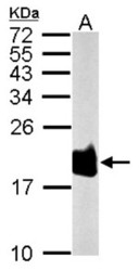
- Western Blot: Superoxide Dismutase 1 Antibody [NBP2-20534] - Sample (50 ug of whole cell lysate) A: Rat brain, 15% SDS PAGE gel, diluted at 1:1000.

- Submitted by
- Novus Biologicals (provider)
- Main image
- Experimental details
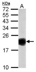
- Western Blot: Superoxide Dismutase 1 Antibody [NBP2-20534] - Sample (50 ug of whole cell lysate) A: Mouse Brain, 15% SDS PAGE gel, diluted at 1:1000.

- Submitted by
- Novus Biologicals (provider)
- Main image

- Experimental details
- Western Blot: SOD1/Cu-Zn SOD Antibody [NBP2-20534] - Non-transfected (-) and transfected (+) 293T whole cell extracts (30 ug) were separated by 15% SDS-PAGE, and the membrane was blotted with SOD1 antibody. HRP-conjugated anti-rabbit IgG antibody was used to detect the primary antibody.
- Submitted by
- Novus Biologicals (provider)
- Main image
- Experimental details
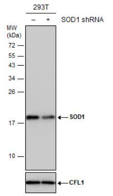
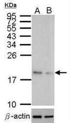
- Western Blot: SOD1/Cu-Zn SOD Antibody [NBP2-20534] - Western blot analysis of SOD1 (upper panel) and beta-actin (lower panel) Sample (10 ug of whole cell lysate) A: HeLa mock control B: HeLa transfected shSOD1 15% SDS PAGE HRP-conjugated anti-rabbit IgG antibody was used to detect the primary antibody.

- Submitted by
- Novus Biologicals (provider)
- Main image
- Experimental details
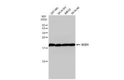
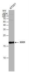
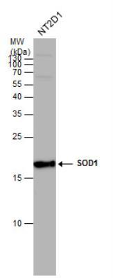
- Western Blot: SOD1/Cu-Zn SOD Antibody [NBP2-20534] - Various whole cell extracts (30 ug) were separated by 15% SDS-PAGE, and the membrane was blotted with SOD1 antibody diluted at 1:1000. HRP-conjugated anti-rabbit IgG antibody was used to detect the primary antibody.
- Submitted by
- Novus Biologicals (provider)
- Main image
- Experimental details
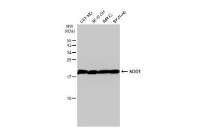
- Western Blot: SOD1/Cu-Zn SOD Antibody [NBP2-20534] - SOD1 antibody detects SOD1 protein by western blot analysis. Whole cell extracts (30 ug) was separated by 15% SDS-PAGE, and the membrane was blotted with SOD1 antibody at a dilution of 1:1000. HRP-conjugated anti-rabbit IgG antibody was used to detect the primary antibody.
Supportive validation
- Submitted by
- Novus Biologicals (provider)
- Main image
- Experimental details
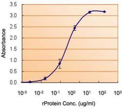
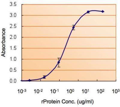
- ELISA: SOD1/Cu-Zn SOD Antibody [NBP2-20534] - ELISA detection of SOD1 using SOD1 Antibody for detection at a concentration of 1.5 ug/mL.
Supportive validation
- Submitted by
- Novus Biologicals (provider)
- Main image

- Experimental details
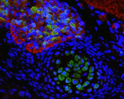
- Immunohistochemistry-Frozen: SOD1/Cu-Zn SOD Antibody [NBP2-20534] - Immunofluorescence photomicrographs of paraffin-embedded mouse fetal brain. Green: SOD1 antibody diluted at 1:200. The signal was developed using goat anti-rabbit IgG antibody (Dylight488). Red: beta Tubulin 3/ TUJ1 antibody diluted at 1:100. The signal was developed using goat anti-mouse IgG antibody (Dylight594). Blue: Nuclear staining with Hoechst 33342. Antigen Retrieval: Citrate buffer, pH 6.0, 15 min
- Submitted by
- Novus Biologicals (provider)
- Main image

- Experimental details
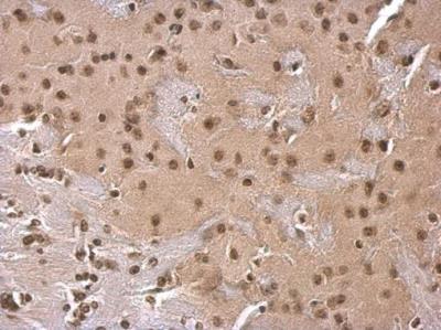
- Immunohistochemistry-Paraffin: SOD1/Cu-Zn SOD Antibody [NBP2-20534] - SOD1 antibody detects SOD1 protein at cytosol on mouse fore brain by immunohistochemical analysis. Sample: Paraffin-embedded mouse fore brain. SOD1 antibody dilution: 1:500.