Explore
 Validate
 Learn
Learn Western blot
Antibody data
- Antibody Data
- Antigen structure
- References [0]
- Comments [0]
- Validations
- Western blot [1]
- Flow cytometry [1]
Submit
Validation data
Reference
Comment
Report error
- Product number
- MAB11241-100 - Provider product page

- Provider
- Novus Biologicals
- Product name
- Rat Monoclonal CD11b Antibody
- Antibody type
- Monoclonal
- Description
- Protein A or G purified from hybridoma culture supernatant. Detects recombinant mouse CD11b/Integrin alpha M in Western blot.
- Reactivity
- Mouse
- Host
- Rat
- Conjugate
- Unconjugated
- Isotype
- IgG
- Vial size
- 100 ug
- Storage
- Use a manual defrost freezer and avoid repeated freeze-thaw cycles. 12 months from date of receipt, -20 to -70 degreesC as supplied. 1 month, 2 to 8 degreesC under sterile conditions after reconstitution. 6 months, -20 to -70 degreesC under sterile conditions after reconstitution.
No comments: Submit comment
Supportive validation
- Submitted by
- Novus Biologicals (provider)
- Main image
- Experimental details
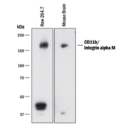
- Detection of Mouse CD11b/Integrin alpha M by Western Blot. Western blot shows lysates of RAW 264.7 mouse monocyte/macrophage cell line. PVDF membrane was probed with 1 µg/mL of Mouse Anti-Mouse CD11b/Integrin alpha M Monoclonal Antibody (Catalog # MAB11241) followed by HRP-conjugated Anti-Rat IgG Secondary Antibody (Catalog # HAF005). A specific band was detected for CD11b/Integrin alpha M at approximately 160 kDa (as indicated). This experiment was conducted under reducing conditions and using Immunoblot Buffer Group 1.

Supportive validation
- Submitted by
- Novus Biologicals (provider)
- Main image

- Experimental details
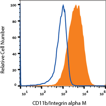
- Detection of CD11b/Integrin alpha M in RAW 264.7 Mouse Cell Line by Flow Cytometry RAW 264.7 mouse monocyte/macrophage cell line was stained with Rat Anti-Mouse CD11b/Integrin alpha M Monoclonal Antibody (Catalog # MAB11241, filled histogram) or isotype control antibody (Catalog # MAB0061, open histogram), followed by Phycoerythrin-conjugated Anti-Rat IgG Secondary Antibody (Catalog # F0105B). View our protocol for Staining Membrane-associated Proteins.