Explore
 Validate
 Learn
Learn Flow cytometry
Antibody data
- Antibody Data
- Antigen structure
- References [1]
- Comments [0]
- Validations
- Flow cytometry [1]
Submit
Validation data
Reference
Comment
Report error
- Product number
- 12-5785-82 - Provider product page

- Provider
- Invitrogen Antibodies
- Product name
- NOTCH1 Monoclonal Antibody (mN1A), PE, eBioscience™
- Antibody type
- Monoclonal
- Antigen
- Other
- Description
- Description: The Notch family of transmembrane receptors controls cell-fate decisions during the development of many organs in a wide variety of species. After binding its ligand, the Notch receptor is cleaved in its transmembrane domain, and the resulting intracellular domain dissociates from the membrane and translocates to the nucleus, where it is able to suppress the expression of lineage-specific genes by interacting with transcriptional repressors. The mN1A antibody reacts with the intracellular domain of mouse and human Notch1, but not with Notch2, 3, or 4. The mN1A antibody has a low affinity for the full-length (unprocessed or heterodimeric cell surface) forms of Notch1. In the mouse, Notch mRNA is expressed in mouse hematopoietic cells of the fetal liver and adult thymus and bone marrow. In the thymus, Notch1 protein is detected in CD4-CD8- (double-negative) and CD4-CD8+ (single-positive) thymocytes. Studies of Notch1-transgenic cells and Notch1-null mice indicate that the receptor is involved in the regulation of lymphopoiesis and myelopoiesis.
- Conjugate
- Yellow dye
- Antibody clone number
- mN1A
- Concentration
- 0.2 mg/mL
Submitted references NOTCH1 Is Aberrantly Activated in Chronic Lymphocytic Leukemia Hematopoietic Stem Cells.
Di Ianni M, Baldoni S, Del Papa B, Aureli P, Dorillo E, De Falco F, Albi E, Varasano E, Di Tommaso A, Giancola R, Accorsi P, Rotta G, Rompietti C, Silva Barcelos EC, Campese AF, Di Bartolomeo P, Screpanti I, Rosati E, Falzetti F, Sportoletti P
Frontiers in oncology 2018;8:105
Frontiers in oncology 2018;8:105
No comments: Submit comment
Supportive validation
- Submitted by
- Invitrogen Antibodies (provider)
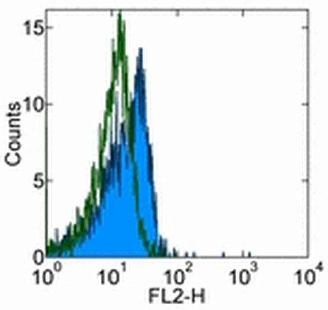
- Main image

- Experimental details
- Intracellular staining of C57BL/6 thymocytes with 0.06 µg of Mouse IgG1 kappa Isotype Control PE (Product # 12-4714-81) (open histogram) or 0.125 µg of Anti-Human/Mouse Notch1 PE (filled histogram) using the Intracellular Fixation & Permeabilization Buffer Set (Product # 88-8824-00) and protocol. CD4-CD8. thymocytes were used for analysis.
- Conjugate
- Yellow dye