Explore
 Validate
 Learn
Learn Western blot
Antibody data
- Antibody Data
- Antigen structure
- References [2]
- Comments [0]
- Validations
- Western blot [2]
Submit
Validation data
Reference
Comment
Report error
- Product number
- AF1149 - Provider product page

- Provider
- R&D Systems
- Product name
- Mouse/Rat Serpin F1/PEDF Antibody
- Antibody type
- Polyclonal
- Description
- Antigen Affinity-purified. Detects mouse Serpin F1/PEDF in direct ELISAs and Western blots. In direct ELISAs, approximately 35% cross-reactivity with recombinant human Serpin F1 is observed.
- Reactivity
- Mouse, Rat
- Host
- Goat
- Conjugate
- Unconjugated
- Antigen sequence
BAA31978- Isotype
- IgG
- Vial size
- 100 ug
- Concentration
- LYOPH
- Storage
- Use a manual defrost freezer and avoid repeated freeze-thaw cycles. 12 months from date of receipt, -20 to -70 °C as supplied. 1 month, 2 to 8 °C under sterile conditions after reconstitution. 6 months, -20 to -70 °C under sterile conditions after reconstitution.
Submitted references Systemic administration of erythropoietin inhibits retinopathy in RCS rats.
Adeno-associated virus vector-mediated delivery of pigment epithelium-derived factor restricts neuroblastoma angiogenesis and growth.
Shen W, Chung SH, Irhimeh MR, Li S, Lee SR, Gillies MC
PloS one 2014;9(8):e104759
PloS one 2014;9(8):e104759
Adeno-associated virus vector-mediated delivery of pigment epithelium-derived factor restricts neuroblastoma angiogenesis and growth.
Streck CJ, Zhang Y, Zhou J, Ng C, Nathwani AC, Davidoff AM
Journal of pediatric surgery 2005 Jan;40(1):236-43
Journal of pediatric surgery 2005 Jan;40(1):236-43
No comments: Submit comment
Supportive validation
- Submitted by
- R&D Systems (provider)
- Main image
- Experimental details
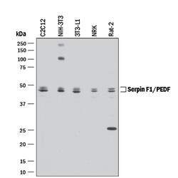
- Detection of Mouse and Rat Serpin F1/PEDF by Western Blot. Western blot shows lysates of C2C12 mouse myoblast cell line, NIH-3T3 mouse embryonic fibroblast cell line, 3T3-L1 mouse embryonic fibroblast adipose-like cell line, NRK rat normal kidney cell line, and Rat-2 rat embryonic fibroblast cell line. PVDF membrane was probed with 0.2 µg/mL of Goat Anti-Mouse/Rat Serpin F1/PEDF Antigen Affinity-purified Polyclonal Antibody (Catalog # AF1149) followed by HRP-conjugated Anti-Goat IgG Secondary Antibody (Catalog # HAF017). Specific bands were detected for Serpin F1/PEDF at approximately 46-48 kDa (as indicated). This experiment was conducted under reducing conditions and using Immunoblot Buffer Group 1.

- Submitted by
- R&D Systems (provider)
- Main image
- Experimental details
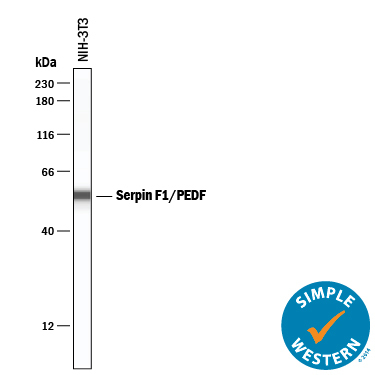
- Detection of Mouse Serpin F1/PEDF by Simple WesternTM. Simple Western lane view shows lysates of NIH-3T3 mouse embryonic fibroblast cell line, loaded at 0.2 mg/mL. A specific band was detected for Serpin F1/PEDF at approximately 55 kDa (as indicated) using 20 µg/mL of Goat Anti-Mouse/Rat Serpin F1/PEDF Antigen Affinity-purified Polyclonal Antibody (Catalog # AF1149) followed by 1:50 dilution of HRP-conjugated Anti-Goat IgG Secondary Antibody (Catalog # HAF109). This experiment was conducted under reducing conditions and using the 12-230 kDa separation system.