Explore
 Validate
 Learn
Learn Western blot
Antibody data
- Antibody Data
- Antigen structure
- References [0]
- Comments [0]
- Validations
- Western blot [1]
- Immunocytochemistry [2]
Submit
Validation data
Reference
Comment
Report error
- Product number
- TA324748 - Provider product page

- Provider
- OriGene
- Product name
- Rabbit polyclonal anti-HSPD1(HSP60) antibody(Center), Loading control
- Antibody type
- Polyclonal
- Description
- Rabbit polyclonal anti-HSPD1(HSP60) antibody(Center), Loading control
- Host
- Rabbit
- Conjugate
- Unconjugated
- Epitope
- HSPD1
- Isotype
- IgG
- Antibody clone number
- NULL
- Vial size
- 400 µl
- Concentration
- 2.0 mg/ml
No comments: Submit comment
Supportive validation
- Submitted by
- OriGene (provider)
- Main image
- Experimental details
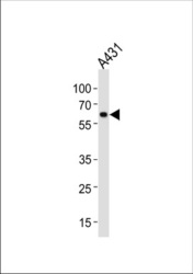
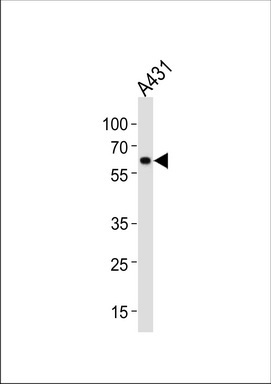
- HSPD1 Antibody (Center) (Cat. #TA324748) western blot analysis in A431 cell line lysates (35ug/lane).This demonstrates the HSPD1 antibody detected the HSPD1 protein (arrow).
- Validation comment
- WB
Supportive validation
- Submitted by
- OriGene (provider)
- Main image

- Experimental details
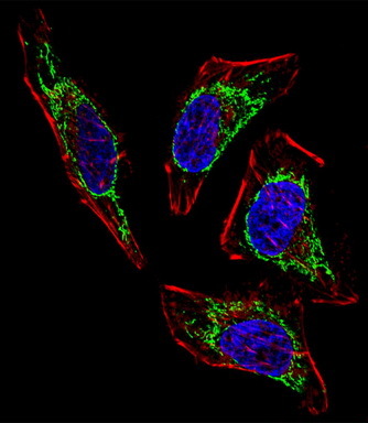
- IF image of U251 cells stained with HSPD1 (Center) antibody. U251 cells were incubated with AP2859C HSPD1 (Center) primary antibody (1:100, 2 h at RT). For secondary antibody, Alexa Fluor? 488 conjugated donkey anti-rabbit antibody (green) was used (1:1000, 1h). Cytoplasmic actin was counterstained with Alexa Fluor? 555 (red) conjugated Phalloidin . Pictures were taken on a Biorevo microscope (BZ-900, Keyence). Note the highly specific localization of the HSPD1 mainly to the mitochondria.
- Validation comment
- IF
- Submitted by
- OriGene (provider)
- Main image
- Experimental details
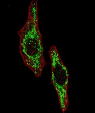
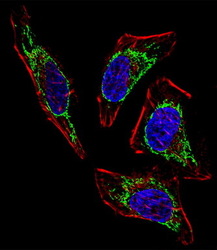
- IF image of Hela cell stained with HSPD1 Antibody (Center)(Cat#TA324748). Hela cells were incubated with HSPD1 primary antibody (1:25, 1 h at 37?). For secondary antibody, Alexa Fluor? 488 conjugated donkey anti-rabbit antibody (green) was used (1:400).Cytoplasmic actin was counterstained with Alexa Fluor? 555 (red) conjugated Phalloidin (7 units/ml). Nuclei were counterstained with DAPI (blue) . HSPD1 immunoreactivity is localized to Mitochondria significantly.
- Validation comment
- IF