Explore
 Validate
 Learn
Learn Western blot
 ELISA
ELISAAntibody data
- Antibody Data
- Antigen structure
- References [0]
- Comments [0]
- Validations
- Western blot [1]
- Immunoprecipitation [1]
- Immunohistochemistry [2]
Submit
Validation data
Reference
Comment
Report error
- Product number
- NB120-5479 - Provider product page

- Provider
- Novus Biologicals
- Proper citation
- Novus Cat#NB120-5479, RRID:AB_790265
- Product name
- Mouse Monoclonal HSP60 Antibody
- Antibody type
- Monoclonal
- Description
- Immunogen affinity purified. Detects Hsp 60 from human cells, tissues and recombinant protein preparations. This displays no other protein or species cross-reactivity.
- Reactivity
- Human, Mouse
- Host
- Mouse
- Antigen sequence
Epitope mapping studies using human
 Hsp 60 deletion mutants suggest th
at this antibody binds between amin
o acids 211-288.- Isotype
- IgM
- Vial size
- 100ug
- Concentration
- LYOPH
- Storage
- Store at -20C. Avoid freeze-thaw cycles.
No comments: Submit comment
Supportive validation
- Submitted by
- Novus Biologicals (provider)
- Main image
- Experimental details
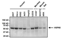
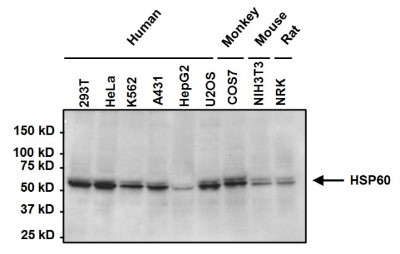
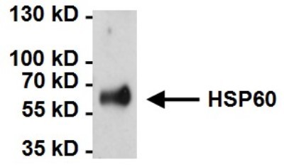
- Western Blot: HSP60 Antibody (2E1/53) [NB120-5479] - Analysis of Heat Shock Protein 60 (HSP 60) was performed by loading 50ug of the indicated whole cell lysates onto a 4-20% Tris-HCl polyacrylamide gel. Proteins were transferred to a PVDF membrane and blocked with 5% BSA/TBST for at least 1 hour. The membrane was probed with a HSP60 monoclonal antibody at a concentration of 1ug/ml overnight at 4C on a rocking platform, washed in TBS-0.1%Tween 20, and probed with a goat anti-mouse IgM secondary antibody at a dilution of 1:20,000 for at least 1 hour. Chemiluminescent detection was performed using SuperSignal West Pico.
Supportive validation
- Submitted by
- Novus Biologicals (provider)
- Main image
- Experimental details
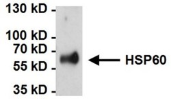
- Immunoprecipitation: HSP60 Antibody (2E1/53) [NB120-5479] - Analysis of Heat Shock Protein 60 (HSP60) was performed on HeLa cells. Antigen-antibody complexes were formed by incubating 500ug of whole cell lysate with 2ug of a HSP60 monoclonal antibody overnight on a rocking platform at 4C. The immune complexes were captured on 50ul Protein A/G Plus Agarose, washed extensively, and eluted with 5X Lane Marker Reducing Sample Buffer. Samples were resolved on a 4-20% Tris-HCl polyacrylamide gel, transferred to a PVDF membrane, and blocked with 5% BSA/TBST for at least 1 hour. The membrane was probed with a HSP60 monoclonal antibody at a concentration of 1ug/ml overnight rotating at 4C, washed in TBST, and probed with a goat anti-mouse IgM secondary antibody at a dilution of 1:20,000 for at least 1 hour. Chemiluminescent detection performed using SuperSignal West Dura.
Supportive validation
- Submitted by
- Novus Biologicals (provider)
- Main image
- Experimental details
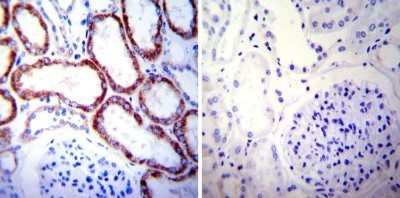
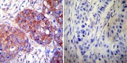
- Immunohistochemistry-Paraffin: HSP60 Antibody (2E1/53) [NB120-5479] - Both normal and cancer biopsies of deparaffinized Human breast carcinoma tissues.

- Submitted by
- Novus Biologicals (provider)
- Main image
- Experimental details
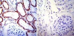
- Immunohistochemistry-Paraffin: HSP60 Antibody (2E1/53) [NB120-5479] - Both normal and cancer biopsies of deparaffinized Human kidney tissue tissues.