Explore
 Validate
 Learn
Learn Western blot
 ELISA
ELISAAntibody data
- Antibody Data
- Antigen structure
- References [0]
- Comments [0]
- Validations
- Western blot [2]
- Immunohistochemistry [1]
Submit
Validation data
Reference
Comment
Report error
- Product number
- PA5-79451 - Provider product page

- Provider
- Invitrogen Antibodies
- Product name
- IGFBP3 Polyclonal Antibody
- Antibody type
- Polyclonal
- Antigen
- Synthetic peptide
- Description
- Reconstitute with 0.2 mL of distilled water to yield a concentration of 500 µg/mL.
- Reactivity
- Human, Mouse, Rat
- Host
- Rabbit
- Isotype
- IgG
- Vial size
- 100 µg
- Concentration
- 500 µg/mL
- Storage
- -20°C
No comments: Submit comment
Supportive validation
- Submitted by
- Invitrogen Antibodies (provider)
- Main image

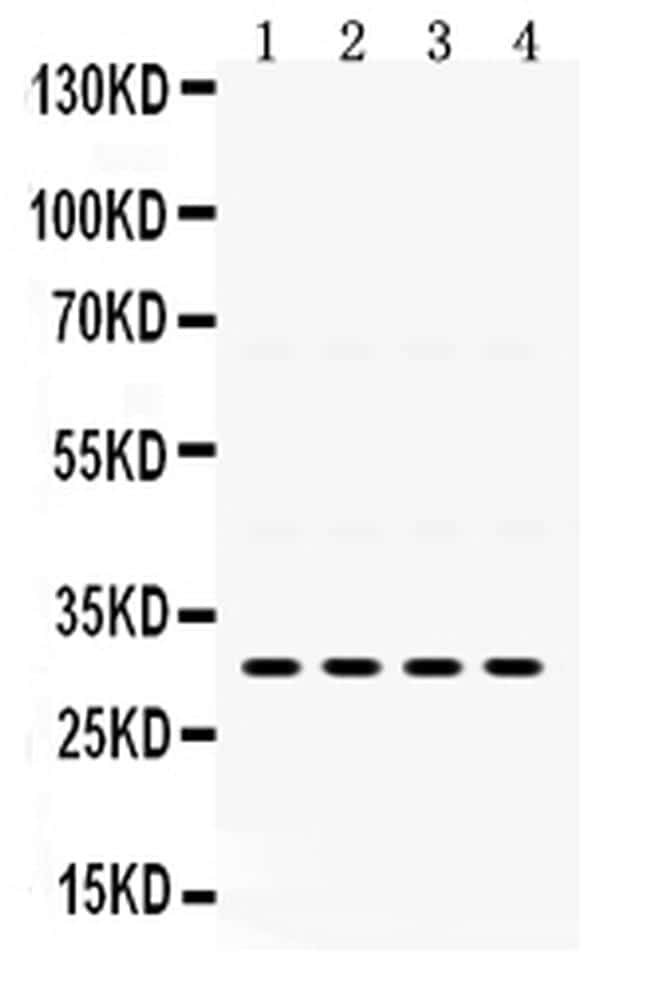
- Experimental details
- Western blot analysis of IGFBP3 in Lane 1: rat kidney tissue lysate, Lane 2: rat liver tissue lysate, Lane 3: SGC whole cell lysate, Lane 4: 22RV1 whole cell lysate using 40-50 µg per well. Sample was incubated with IGFBP3 (Product # PA5-79451) at a dilution of 0.5 µg/mL.
- Submitted by
- Invitrogen Antibodies (provider)
- Main image
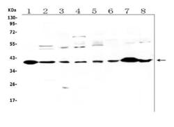
- Experimental details
- Western blot analysis of IGFBP-3 in, Lane 1: rat heart tissue lysates, Lane 2: rat brain tissue lysates, Lane 3: rat liver tissue lysates, Lane 4: rat PC-12 whole cell lysates. Lane 5: human U-87MG whole cell lysates. Lane 6: mouse kidney tissue lysates, Lane 7: mouse heart tissue lysates, Lane 8: mouse brain tissue lysates, . Electrophoresis was performed on a 5-20% SDS-PAGE gel at 70V (Stacking gel) / 90V (Resolving gel) for 2-3 hours. The sample well of each lane was loaded with 50 µg of sample under reducing conditions. After Electrophoresis, proteins were transferred to a Nitrocellulose membrane at 150mA for 50-90 minutes. The membrane was blocked with 5% Non-fat Milk/ TBS for 1. 5 hour at RT. The membrane was incubated with IGFBP3 Polyclonal Antibody (Product # PA5-79451) at 0.5 μg/mL overnight at 4°C, then washed with TBS-0. 1% Tween 3 times with 5 minutes each and probed with a goat anti-rabbit IgG-HRP secondary antibody at a dilution of 1:10000 for 1. 5 hour at RT. The signal is developed using an Enhanced Chemiluminescent detection (ECL) kit. A specific band was detected for IGFBP-3 at approximately 40KD. The expected band size for IGFBP-3 is at 32KD.

Supportive validation
- Submitted by
- Invitrogen Antibodies (provider)
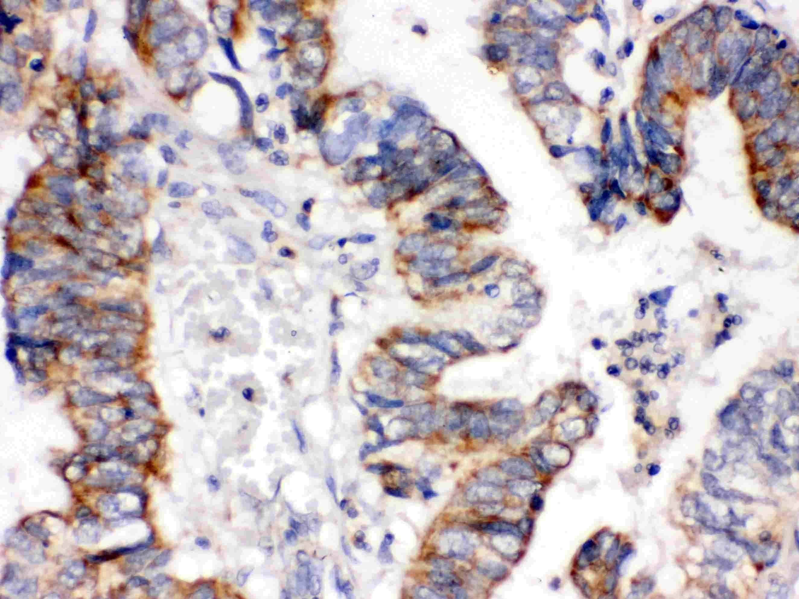
- Main image
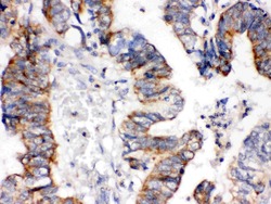
- Experimental details
- Immunohistochemistry analysis of IGFBP3 on paraffin-embedded human intestinal cancer tissue. Sample was incubated with IGFBP3 polyclonal antibody (Product# PA5-79451).