Explore
 Validate
 Learn
Learn ELISA
ELISAAntibody data
- Antibody Data
- Antigen structure
- References [3]
- Comments [0]
- Validations
- ELISA [1]
- Immunohistochemistry [1]
- Flow cytometry [1]
Submit
Validation data
Reference
Comment
Report error
- Product number
- MAB941-100 - Provider product page

- Provider
- R&D Systems
- Product name
- Human SPARC Antibody
- Antibody type
- Monoclonal
- Description
- Protein A or G purified from hybridoma culture supernatant. Detects human SPARC/Osteonectin in direct ELISAs. In direct ELISAs, no cross-reactivity with recombinant mouse SPARC/Osteonectin is observed.
- Reactivity
- Human
- Host
- Mouse
- Conjugate
- Unconjugated
- Antigen sequence
P09486- Isotype
- IgG
- Antibody clone number
- 122511
- Vial size
- 100 ug
- Storage
- Use a manual defrost freezer and avoid repeated freeze-thaw cycles. 12 months from date of receipt, -20 to -70 °C as supplied. 1 month, 2 to 8 °C under sterile conditions after reconstitution. 6 months, -20 to -70 °C under sterile conditions after reconstitution.
Submitted references Potential actionable targets in appendiceal cancer detected by immunohistochemistry, fluorescent in situ hybridization, and mutational analysis.
Insulin-like growth factor binding protein-4 (IGFBP-4) is a novel anti-angiogenic and anti-tumorigenic mediator secreted by dibutyryl cyclic AMP (dB-cAMP)-differentiated glioblastoma cells.
Biomarker discovery from pancreatic cancer secretome using a differential proteomic approach.
Borazanci E, Millis SZ, Kimbrough J, Doll N, Von Hoff D, Ramanathan RK
Journal of gastrointestinal oncology 2017 Feb;8(1):164-172
Journal of gastrointestinal oncology 2017 Feb;8(1):164-172
Insulin-like growth factor binding protein-4 (IGFBP-4) is a novel anti-angiogenic and anti-tumorigenic mediator secreted by dibutyryl cyclic AMP (dB-cAMP)-differentiated glioblastoma cells.
Moreno MJ, Ball M, Andrade MF, McDermid A, Stanimirovic DB
Glia 2006 Jun;53(8):845-57
Glia 2006 Jun;53(8):845-57
Biomarker discovery from pancreatic cancer secretome using a differential proteomic approach.
Grønborg M, Kristiansen TZ, Iwahori A, Chang R, Reddy R, Sato N, Molina H, Jensen ON, Hruban RH, Goggins MG, Maitra A, Pandey A
Molecular & cellular proteomics : MCP 2006 Jan;5(1):157-71
Molecular & cellular proteomics : MCP 2006 Jan;5(1):157-71
No comments: Submit comment
Supportive validation
- Submitted by
- R&D Systems (provider)
- Main image

- Experimental details
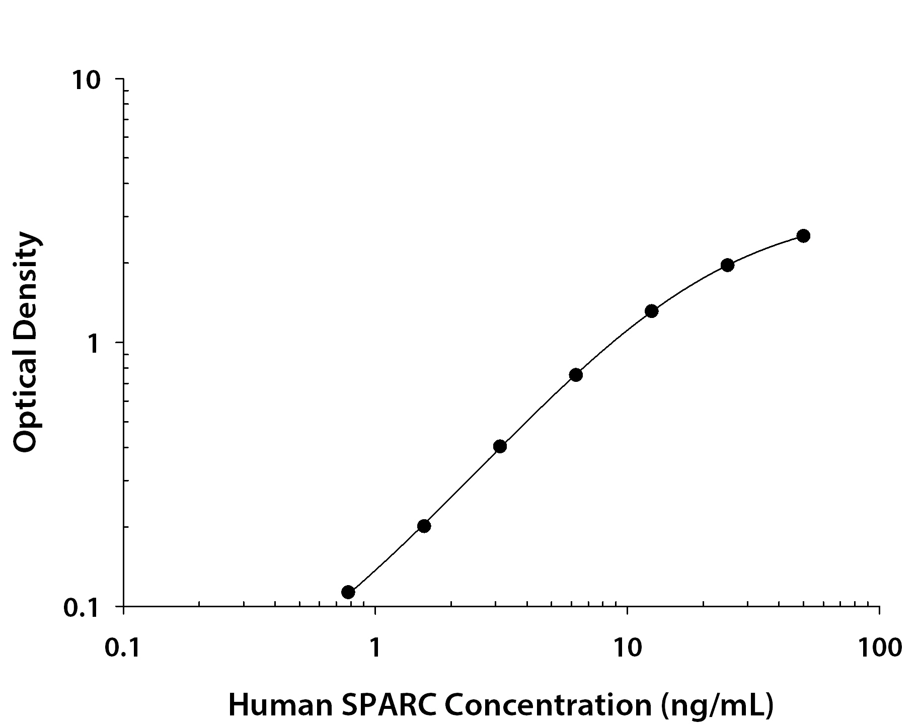
- Human SPARC ELISA Standard Curve. Recombinant Human VSIG8 protein was serially diluted 2-fold and captured by Mouse Anti-Human VSIG8 Monoclonal Antibody (Catalog # MAB9418) coated on a Clear Polystyrene Microplate (Catalog # DY990). Goat Anti-Human SPARC Antigen Affinity-purified Polyclonal Antibody (Catalog # AF941) was biotinylated and incubated with the protein captured on the plate. Detection of the standard curve was achieved by incubating Streptavidin-HRP (Catalog # DY998) followed by Substrate Solution (Catalog # DY999) and stopping the enzymatic reaction with Stop Solution (Catalog # DY994).
Supportive validation
- Submitted by
- R&D Systems (provider)
- Main image

- Experimental details
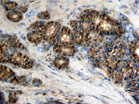
- SPARC/Osteonectin in Human Ovary Cancer Tissue. SPARC/Osteonectin was detected in immersion fixed paraffin-embedded sections of human ovarian clear cell carcinoma tissue using Human SPARC/Osteonectin Monoclonal Antibody (Catalog # MAB941) at 25 µg/mL overnight at 4 °C. Tissue was stained using the Anti-Mouse HRP-DAB Cell & Tissue Staining Kit (brown; Catalog # CTS002) and counterstained with hematoxylin (blue). View our protocol for Chromogenic IHC Staining of Paraffin-embedded Tissue Sections.
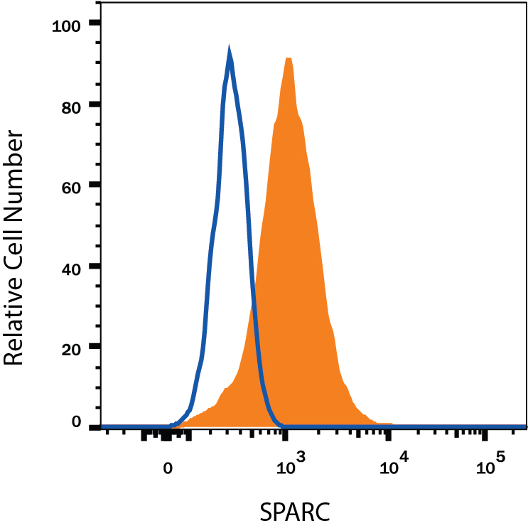
Supportive validation
- Submitted by
- R&D Systems (provider)
- Main image

- Experimental details
- Detection of SPARC in HT1080 Human Cell Line by Flow Cytometry. HT1080 human fibrosarcoma cell line was stained with Mouse Anti-Human SPARC Monoclonal Antibody (Catalog # MAB941, filled histogram) or isotype control antibody (Catalog # MAB002, open histogram), followed by Allophycocyanin-conjugated Anti-Mouse IgG Secondary Antibody (Catalog # F0101B). To facilitate intracellular staining, cells were fixed with Flow Cytometry Fixation Buffer (Catalog # FC004) and permeabilized with Flow Cytometry Permeabilization/Wash Buffer I (Catalog # FC005). View our protocol for Staining Intracellular Molecules.