Explore
 Validate
 Learn
Learn Western blot
 Immunohistochemistry
Antibody data
- Antibody Data
- Antigen structure
- References [0]
- Comments [0]
- Validations
- Western blot [2]
Submit
Validation data
Reference
Comment
Report error
- Product number
- PA1-26253 - Provider product page

- Provider
- Invitrogen Antibodies
- Product name
- Cathepsin D Polyclonal Antibody
- Antibody type
- Polyclonal
- Antigen
- Other
- Description
- Recommended positive controls: Breast carcinoma.
- Reactivity
- Human
- Host
- Rabbit
- Isotype
- IgG
- Vial size
- 250 µL
- Storage
- 4° C, do not freeze
No comments: Submit comment
Supportive validation
- Submitted by
- Invitrogen Antibodies (provider)
- Main image

- Experimental details
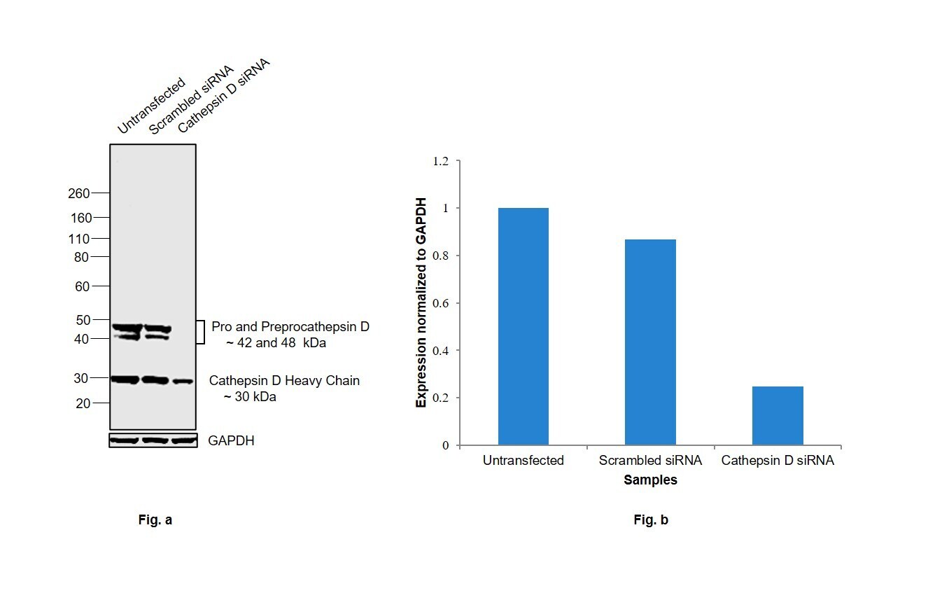
- Knockdown of Cathepsin D was achieved by transfecting MCF7 with Cathepsin D specific siRNAs (Silencer® select Product # S137, S136). Western blot analysis (Fig. a) was performed using Whole cell extracts from the Cathepsin D knockdown cells (lane 3), non-targeting scrambled siRNA transfected cells (lane 2) and untransfected cells (lane 1). The blot was probed with Cathepsin D Polyclonal Antibody (Product # PA1-26253, 1:1000 dilution ) and Goat anti-Rabbit IgG (H+L) Superclonal™ Recombinant Secondary Antibody, HRP (Product # A27036, 1:4000 dilution). Densitometric analysis of this western blot is shown in histogram (Fig. b). Decrease in signal upon siRNA mediated knock down confirms that antibody is specific to Cathepsin D.
- Submitted by
- Invitrogen Antibodies (provider)
- Main image
- Experimental details
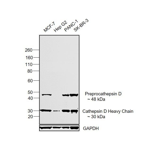
- Western blot was performed using Anti-Cathepsin D Polyclonal Antibody (Product # PA1-26253) and a 48 kDa band corresponding to Preprocathepsin D along with a 30 kDa band that corresponds to Cathepsin D heavy chain was observed across cell lines tested. Whole cell extracts (30 µg lysate) of MCF7 (Lane 1), Hep G2 (Lane 2), PANC-1 (Lane 3) and SK-BR-3 (Lane 4) were electrophoresed using NuPAGE™ 4-12% Bis-Tris Protein Gel (Product # NP0322BOX). Resolved proteins were then transferred onto a Nitrocellulose membrane (Product # IB23001) by iBlot® 2 Dry Blotting System (Product # IB21001). The blot was probed with the primary antibody (1:1000 dilution) and detected by chemiluminescence with Goat anti-Rabbit IgG (H+L) Superclonal™ Recombinant Secondary Antibody, HRP (Product # A27036, 1:4000 dilution) using the iBright FL 1000 (Product # A32752). Chemiluminescent detection was performed using Novex® ECL Chemiluminescent Substrate Reagent Kit (Product # WP20005).