 Explore
 Validate
 Learn
Learn Western blot
Antibody data
- Antibody Data
- Antigen structure
- References [1]
- Comments [0]
- Validations
- Western blot [2]
- Immunohistochemistry [1]
Submit
Validation data
Reference
Comment
Report error
- Product number
- MAB1014 - Provider product page

- Provider
- Novus Biologicals
- Product name
- Mouse Monoclonal Cathepsin D Antibody
- Antibody type
- Monoclonal
- Description
- Protein A or G purified from hybridoma culture supernatant. Detects human Cathepsin D in direct ELISAs and Western blots. It recognizes both the pro and mature forms of recombinant human (rh) Cathepsin D. In direct ELISAs, no cross-reactivity with rhCathepsin A, rhCathepsin B, rhCathepsin C, rhCathepsin L, rhCathepsin O, rhCathepsin S, rhCathepsin Z, or recombinant mouse Cathepsin D is observed. In Western blots, 100% cross-reactivity with rhCathepsin E and rmCathepsin D is observed and no cross-reactivity with rhBACE-1 is observed.
- Reactivity
- Human
- Host
- Mouse
- Conjugate
- Unconjugated
- Isotype
- IgG
- Vial size
- 500 ug
- Concentration
- LYOPH
- Storage
- Use a manual defrost freezer and avoid repeated freeze-thaw cycles. 12 months from date of receipt, -20 to -70 degreesC as supplied. 1 month, 2 to 8 degreesC under sterile conditions after reconstitution. 6 months, -20 to -70 degreesC under sterile conditions after reconstitution.
Submitted references Biomarker discovery from pancreatic cancer secretome using a differential proteomic approach.
Grønborg M, Kristiansen TZ, Iwahori A, Chang R, Reddy R, Sato N, Molina H, Jensen ON, Hruban RH, Goggins MG, Maitra A, Pandey A
Molecular & cellular proteomics : MCP 2006 Jan;5(1):157-71
Molecular & cellular proteomics : MCP 2006 Jan;5(1):157-71
No comments: Submit comment
Supportive validation
- Submitted by
- Novus Biologicals (provider)
- Main image
- Experimental details
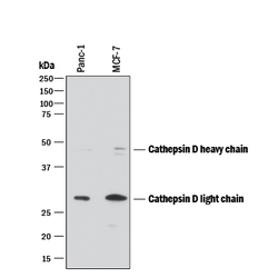
- Detection of Human Cathepsin D by Western Blot. Western blot shows lysates of PANC-1 human pancreatic carcinoma cell line and MCF-7 human breast cancer cell line. PVDF membrane was probed with 0.2 µg/mL of Mouse Anti-Human Cathepsin D Monoclonal Antibody (Catalog # MAB1014) followed by HRP-conjugated Anti-Mouse IgG Secondary Antibody (Catalog # HAF018). Specific bands were detected for Cathepsin D at approximately 28 and 46 kDa (as indicated). This experiment was conducted under reducing conditions and using Immunoblot Buffer Group 1.

- Submitted by
- Novus Biologicals (provider)
- Main image
- Experimental details
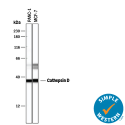
- Detection of Human Cathepsin D by Simple WesternTM. Simple Western lane view shows lysates of PANC-1 human pancreatic carcinoma cell line and MCF-7 human breast cancer cell line, loaded at 0.2 mg/mL. Specific bands were detected for Cathepsin D at approximately 36 and 52-57 kDa (as indicated) using 2 µg/mL of Mouse Anti-Human Cathepsin D Monoclonal Antibody (Catalog # MAB1014) . This experiment was conducted under reducing conditions and using the 12-230 kDa separation system.

Supportive validation
- Submitted by
- Novus Biologicals (provider)
- Main image
- Experimental details
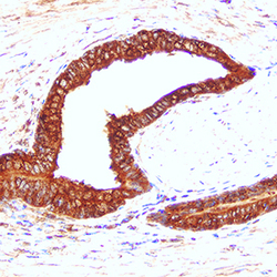
- Cathepsin D in Human Prostate Cancer Tissue. Cathepsin D in Human Prostate Cancer Tissue.
