 Explore
 Validate
 Learn
Learn Western blot
Antibody data
- Antibody Data
- Antigen structure
- References [2]
- Comments [0]
- Validations
- Western blot [9]
- Immunocytochemistry [4]
- Immunohistochemistry [2]
Submit
Validation data
Reference
Comment
Report error
- Product number
- PA5-29688 - Provider product page

- Provider
- Invitrogen Antibodies
- Product name
- Vinculin Polyclonal Antibody
- Antibody type
- Polyclonal
- Antigen
- Recombinant protein fragment
- Description
- Recommended positive controls: NT2D1, Mouse brain, HeLa.
- Concentration
- 0.21 mg/mL
Submitted references Switching to a Standard Chow Diet at Weaning Improves the Effects of Maternal and Postnatal High-Fat and High-Sucrose Diet on Cardiometabolic Health in Adult Male Mouse Offspring.
Fasting improves therapeutic response in hepatocellular carcinoma through p53-dependent metabolic synergism.
Chiñas Merlin A, Gonzalez K, Mockler S, Perez Y, Jia UA, Chicco AJ, Ullevig SL, Chung E
Metabolites 2022 Jun 18;12(6)
Metabolites 2022 Jun 18;12(6)
Fasting improves therapeutic response in hepatocellular carcinoma through p53-dependent metabolic synergism.
Krstic J, Reinisch I, Schindlmaier K, Galhuber M, Riahi Z, Berger N, Kupper N, Moyschewitz E, Auer M, Michenthaler H, Nössing C, Depaoli MR, Ramadani-Muja J, Usluer S, Stryeck S, Pichler M, Rinner B, Deutsch AJA, Reinisch A, Madl T, Chiozzi RZ, Heck AJR, Huch M, Malli R, Prokesch A
Science advances 2022 Jan 21;8(3):eabh2635
Science advances 2022 Jan 21;8(3):eabh2635
No comments: Submit comment
Supportive validation
- Submitted by
- Invitrogen Antibodies (provider)
- Main image
- Experimental details
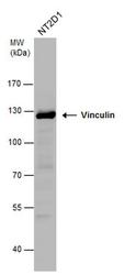
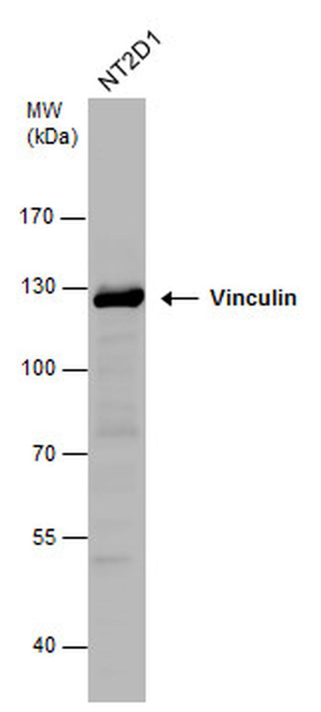
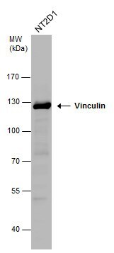
- Western blot analysis of Vinculin using 30 µg of NT2D1 lysate. Samples were loaded onto a 7.5% SDS-PAGE gel and probed with a Vinculin polyclonal antibody (Product # PA5-29688) at a dilution of 1:1000.
- Submitted by
- Invitrogen Antibodies (provider)
- Main image

- Experimental details
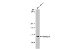
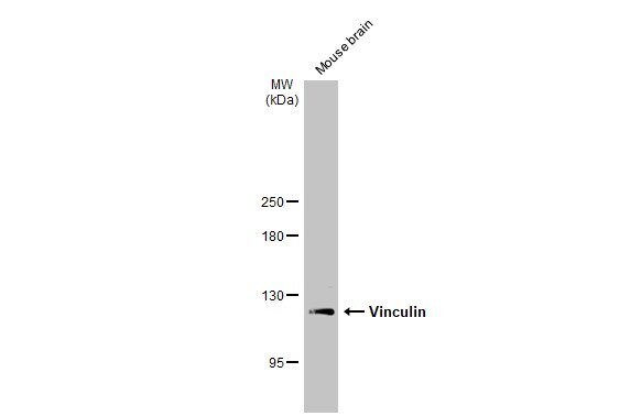
- Western blot analysis of Vinculin using 50 µg of mouse brain lysate. Samples were loaded onto a 7.5% SDS-PAGE gel and probed with a Vinculin polyclonal antibody (Product # PA5-29688) at a dilution of 1:1000.

- Submitted by
- Invitrogen Antibodies (provider)
- Main image
- Experimental details
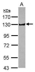
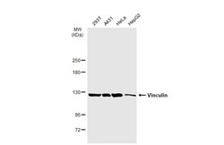
- Western blot analysis of Vinculin in whole cell extracts (30 µg). Samples was separated by 7.5% SDS-PAGE and the membrane was probed with Vinculin Polyclonal antibody (Product # PA5-29688) at a dilution of 1:1000.
- Submitted by
- Invitrogen Antibodies (provider)
- Main image
- Experimental details
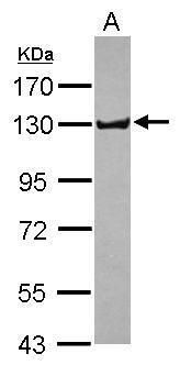
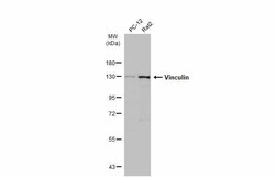
- Western Blot analysis of Vinculin was performed by separating 30 µg of various whole cell extracts by 7.5% SDS-PAGE. Proteins were transferred to a membrane and probed with a Vinculin Polyclonal Antibody (Product # PA5-29688) at a dilution of 1:1000 and a HRP-conjugated anti-rabbit IgG secondary antibody.

- Submitted by
- Invitrogen Antibodies (provider)
- Main image

- Experimental details
- Knockout of Vinculin was achieved by CRISPR-Cas9 genome editing using LentiArray™ Lentiviral sgRNA (Product # A32042, Assay ID CRISPR948199_LV) and LentiArray Cas9 Lentivirus (Product # A32064). Western blot analysis of Vinculin was performed by loading 30 µg of HeLa Wild Type (Lane 1), HeLa Cas9 (Lane 2) andHeLa Vinculin KO (Lane 3) membrane enriched extracts. The samples were electrophoresed using NuPAGE™ Novex™ 4-12% Bis-Tris Protein Gel (Product # NP0322BOX). Resolved proteins were then transferred onto a nitrocellulose membrane (Product # IB23001) by iBlot® 2 Dry Blotting System (Product # IB21001). The blot was probed with Anti-Vinculin Polyclonal Antibody (Product # PA5-29688, 1:2,500 dilution) and Goat anti-Rabbit IgG (H+L) Superclonal™ Recombinant Secondary Antibody, HRP (Product # A27036, 1:6,000 dilution) using the iBright FL 1000 (Product # A32752). Chemiluminescent detection was performed using Novex® ECL Chemiluminescent Substrate Reagent Kit (Product # WP20005). Loss of signal upon CRISPR mediated knockout (KO) using the LentiArray™ CRISPR product line confirms that antibody is specific to Vinculin.

- Submitted by
- Invitrogen Antibodies (provider)
- Main image
- Experimental details
- Western Blot using Vinculin Polyclonal Antibody (Product # PA5-29688). Various whole cell extracts (30 µg) were separated by 5% SDS-PAGE, and the membrane was blotted with Vinculin Polyclonal Antibody (Product # PA5-29688) diluted at 1:1,000. The HRP-conjugated anti-rabbit IgG antibody was used to detect the primary antibody.

- Submitted by
- Invitrogen Antibodies (provider)
- Main image

- Experimental details
- Vinculin antibody detects Vinculin protein by western blot analysis. Whole cell extracts (30 µg) was separated by 7.5% SDS-PAGE, and the membrane was blotted with Vinculin antibody Vinculin Polyclonal Antibody (Product # PA5-29688) diluted at 1:1,000. The HRP-conjugated anti-rabbit IgG antibody was used to detect the primary antibody.
- Submitted by
- Invitrogen Antibodies (provider)
- Main image
- Experimental details
- Western Blot using Vinculin Polyclonal Antibody (Product # PA5-29688). Mouse tissue extract (50 µg) was separated by 5% SDS-PAGE, and the membrane was blotted with Vinculin Polyclonal Antibody (Product # PA5-29688) diluted at 1:1,000. The HRP-conjugated anti-rabbit IgG antibody was used to detect the primary antibody.
- Submitted by
- Invitrogen Antibodies (provider)
- Main image
- Experimental details
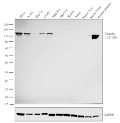
- Western blot analysis was performed on whole cell extracts (30 µg lysate) of HeLa (Lane 1), A-431 (Lane 2), Hep G2 (Lane 3), COS7 (Lane 4), NIH/3T3 (Lane 5), MOLT4 (Lane 6), Ramos (Lane 7), Jurkat (Lane 8), tissue extracts of Mouse liver (Lane 9), Mouse brain (Lane 10) and Human platelets (Lane 11). The blot was probed with Anti-Vinculin Polyclonal Antibody (Product # PA5-29688, 1:2500 dilution) and detected by chemiluminescence using Goat anti-Rabbit IgG (H+L) Superclonal™ Secondary Antibody, HRP conjugate (Product # A27036, 0.25 µg/ml, 1:4000 dilution). A 123 kDa band corresponding to Vinculin was observed across all the cell lines positive for Vinculin (Lanes 1-5) while this band was absent in the cell lines which do not express Vinculin protein (Lanes 6-8). This band was absent in Mouse Liver (Lane 9) while it was present in Mouse Brain (Lane 10) and Human Platelets (Lane 11).

Supportive validation
- Submitted by
- Invitrogen Antibodies (provider)
- Main image

- Experimental details
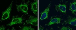
- Immunofluorescent analysis of Vinculin in MCF7 cells fixed in 4% paraformaldehyde at RT for 15 min, using a Vinculin polyclonal antibody (Product # PA5-29688). Green: Primary antibody at a dilution of 1:500. Blue: Hoechst 33342 staining.

- Submitted by
- Invitrogen Antibodies (provider)
- Main image
- Experimental details
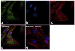
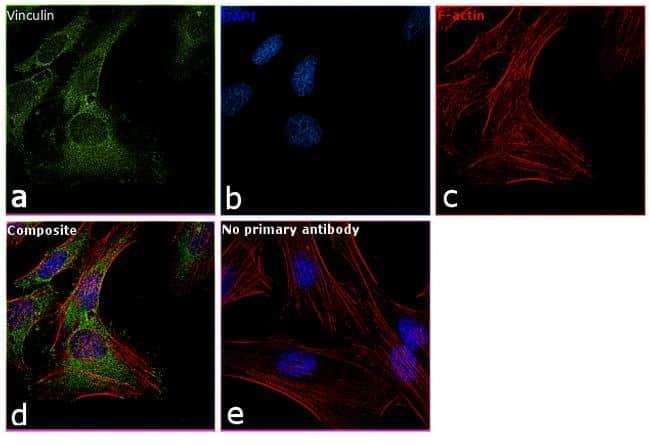
- Immunofluorescence analysis of Vinculin was performed using 70% confluent log phase HeLa cells. The cells were fixed with 4% paraformaldehyde for 10 minutes, permeabilized with 0.1% Triton™ X-100 for 10 minutes, and blocked with 1% BSA for 1 hour at room temperature. The cells were labeled with Vinculin Polyclonal Antibody (Product # PA5-29688) at 1 µg/mL in 0.1% BSA and incubated overnight at 4 degree Celsius and then labeled with Goat anti-Rabbit IgG (H+L) Superclonal™ Secondary Antibody, Alexa Fluor® 488 conjugate (Product # A27034) at a dilution of 1:2000 for 45 minutes at room temperature (Panel a: green). Nuclei (Panel b: blue) were stained with SlowFade® Gold Antifade Mountant with DAPI (Product # S36938). F-actin (Panel c: red) was stained with Rhodamine Phalloidin (Product # R415, 1:300). Panel d represents the merged image showing membranous and cytoplasmic localization. Panel e represents control cells with no primary antibody to assess background. The images were captured at 60X magnification.
- Submitted by
- Invitrogen Antibodies (provider)
- Main image
- Experimental details
- Immunocytochemistry-Immunofluorescence analysis of Vinculin was performed in HeLa cells fixed in ice-cold MeOH for 5 min. Green: Vinculin Polyclonal Antibody (Product # PA5-29688) diluted at 1:500. Blue: Hoechst 33342 staining.

- Submitted by
- Invitrogen Antibodies (provider)
- Main image

- Experimental details
- Vinculin Polyclonal Antibody detects Vinculin protein at focal adhesion by immunofluorescent analysis. Sample: HeLa cells were fixed in ice-cold MeOH for 5 min. Green: Vinculin stained by Vinculin Polyclonal Antibody (Product # PA5-29688) diluted at 1:500. Blue: Fluoroshield with DAPI . Scale bar= 10 µm.

Supportive validation
- Submitted by
- Invitrogen Antibodies (provider)
- Main image
- Experimental details
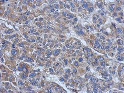
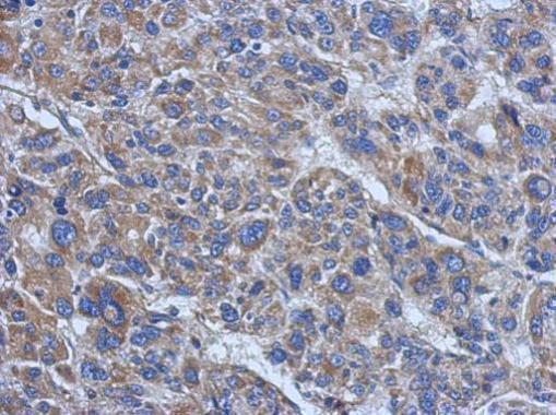
- Immunohistochemical analysis of paraffin-embedded human hepatoma, using Vinculin (Product # PA5-29688) antibody at 1:500 dilution. Antigen Retrieval: EDTA based buffer, pH 8.0, 15 min.
- Submitted by
- Invitrogen Antibodies (provider)
- Main image
- Experimental details
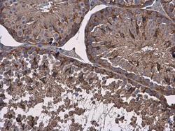
- Vinculin Polyclonal Antibody detects Vinculin protein at cytoplasm in mouse testis by immunohistochemical analysis. Sample: Paraffin-embedded mouse testis. Vinculin Polyclonal Antibody (Product # PA5-29688) diluted at 1:500. Antigen Retrieval: Citrate buffer, pH 6.0, 15 min.
