 Explore
 Validate
 Learn
Learn Western blot
Antibody data
- Antibody Data
- Antigen structure
- References [0]
- Comments [0]
- Validations
- Western blot [1]
- Immunocytochemistry [2]
Submit
Validation data
Reference
Comment
Report error
- Product number
- PA5-66799 - Provider product page

- Provider
- Invitrogen Antibodies
- Product name
- TCF12 Polyclonal Antibody
- Antibody type
- Polyclonal
- Antigen
- Recombinant full-length protein
- Description
- Immunogen sequence: FSPPVNSGKTR PTTLGSSQFS GSGIDERGGT TSWGTSGQPS PSYDSSRGFT DSPHYSDHLN DSRLGAHEGL SPTPFMNSNL MGKTSERGSF SLYSRDTGLP GCQSSLLRQD LGLGSPAQLS SSGKPGTAYY SFSATSSRRR P
- Concentration
- 0.05 mg/mL
No comments: Submit comment
Supportive validation
- Submitted by
- Invitrogen Antibodies (provider)
- Main image
- Experimental details
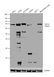
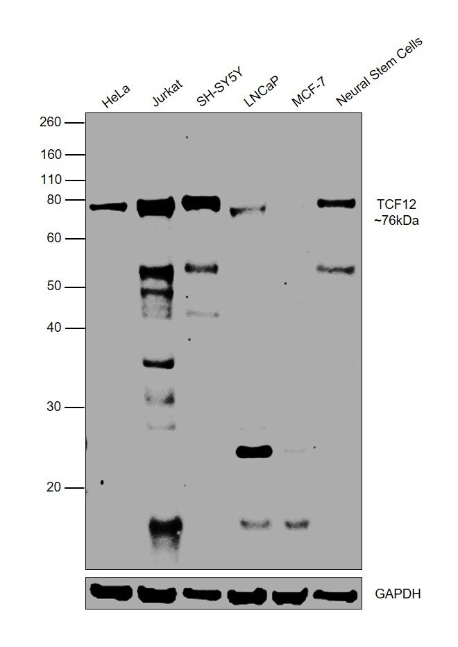
- Western blot was performed using Anti-TCF12 Polyclonal Antibody(Product # PA5-66799) and a 76kDa band corresponding to TCF12 was observed across all tested cell lines, with lower levels in LNCaP and MCF7. Nuclear enriched extracts (30 µg lysate) of HeLa (Lane 1), Jurkat (Lane 2), SH-SY5Y (Lane 3), LNCaP (Lane 4), MCF7 (Lane 5), NSC (Lane 6) were electrophoresed using NuPAGE™ 10% Bis-Tris Protein Gel (Product # NP0302BOX). Resolved proteins were then transferred onto a Nitrocellulose membrane (Product # IB23001) by iBlot® 2 Dry Blotting System (Product # IB21001). The blot was probed with the primary antibody (1 µg/mL) and detected by chemiluminescence with Goat anti-Rabbit IgG (H+L) Superclonal™ Recombinant Secondary Antibody, HRP (Product # A27036,1/4000) using the iBright FL 1000 (Product # A32752). Chemiluminescent detection was performed using Novex® ECL Chemiluminescent Substrate Reagent Kit (Product # WP20005).
Supportive validation
- Submitted by
- Invitrogen Antibodies (provider)
- Main image
- Experimental details
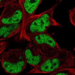
- Immunofluorescent staining of TCF12 in human cell line RH-30 shows localization to nucleoplasm. Samples were probed using a TCF12 Polyclonal Antibody (Product # PA5-66799).

- Submitted by
- Invitrogen Antibodies (provider)
- Main image
- Experimental details
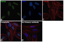
- Immunofluorescence analysis of TCF12 was performed using 70 percent confluent log phase HeLa cells. The cells were fixed with 4% paraformaldehyde for 10 minutes, permeabilized with 0.1% Triton™ X-100 for 15 minutes, and blocked with 2% BSA for 45 minutes at room temperature. The cells were labeled with TCF12 Polyclonal Antibody (Product # PA5-66799) at 5µg/mL in 0.1% BSA, incubated at 4 degree celsius overnight and then labeled with Goat anti-Rabbit IgG (H+L) Highly Cross-Adsorbed Secondary Antibody, Alexa Fluor Plus 488 (Product # A32731), (1:2000), for 45 minutes at room temperature (Panel a: Green). Nuclei (Panel b:Blue) were stained with ProLong™ Diamond Antifade Mountant with DAPI (Product # P36962). F-actin (Panel c: Red) was stained with Rhodamine Phalloidin (Product # R415, 1:300). Panel d represents the merged image showing nuclear localization. Panel e represents control cells with no primary antibody to assess background. The images were captured at 60X magnification.
