Explore
 Validate
 Learn
Learn Western blot
Antibody data
- Antibody Data
- Antigen structure
- References [0]
- Comments [0]
- Validations
- Western blot [2]
- Immunocytochemistry [1]
- Immunohistochemistry [1]
Submit
Validation data
Reference
Comment
Report error
- Product number
- GTX128795 - Provider product page

- Provider
- GeneTex
- Product name
- HIF1 beta antibody
- Antibody type
- Polyclonal
- Reactivity
- Human, Mouse
- Host
- Rabbit
No comments: Submit comment
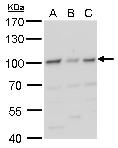
Supportive validation
- Submitted by
- GeneTex (provider)
- Main image

- Experimental details
- HIF1 beta antibody detects HIF1 beta protein by western blot analysis.A. 30 £gg 293T whole cell lysate/extractB. 30 £gg A431whole cell lysate/extractC. 30 £gg HeLa whole cell lysate/extract7.5 % SDS-PAGEHIF1 beta antibody (GTX128795) dilution: 1:1000
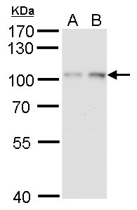
- Submitted by
- GeneTex (provider)
- Main image

- Experimental details
- HIF1 beta antibody detects HIF1 beta protein by western blot analysis.A. 30 £gg HepG2 whole cell lysate/extract (untreated)B. 30 £gg HepG2 whole cell lysate/extract ( 1% O2 treatment for 24 hr)7.5 % SDS-PAGEHIF1 beta antibody (GTX128795) dilution: 1:1000
Supportive validation
- Submitted by
- GeneTex (provider)
- Main image
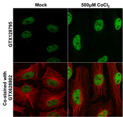
- Experimental details
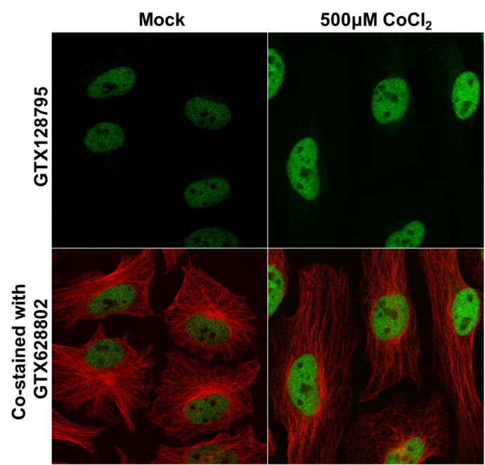
- HIF1 beta antibody detects HIF1 beta protein at nucleus by immunofluorescent analysis. Samples: HeLa cells mock (left) and treated with 500£gM CoCl2 for 24hr (right) were fixed in 4% paraformaldehyde at RT for 15 min.Green: HIF1 beta protein stained by HIF1 beta antibody (GTX128795) diluted at 1:500.Red: alpha Tubulin, a cytoskeleton marker, stained by alpha Tubulin antibody [GT114] (GTX628802) diluted at 1:1000.
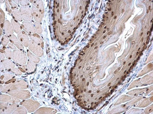
Supportive validation
- Submitted by
- GeneTex (provider)
- Main image

- Experimental details
- HIF1 beta antibody detects HIF1 beta protein at nucleus on mouse esophagus by immunohistochemical analysis. Sample: Paraffin-embedded mouse esophagus. HIF1 beta antibody (GTX128795) dilution: 1:500.