 Explore
 Validate
 Learn
Learn Western blot
Antibody data
- Antibody Data
- Antigen structure
- References [1]
- Comments [0]
- Validations
- Western blot [1]
Submit
Validation data
Reference
Comment
Report error
- Product number
- AF6474 - Provider product page

- Provider
- Novus Biologicals
- Product name
- Sheep Polyclonal FLI1 Antibody
- Antibody type
- Polyclonal
- Description
- Immunogen affinity purified. Detects human FLI1 in Western blots.
- Reactivity
- Human
- Host
- Sheep
- Isotype
- IgG
- Vial size
- 100 ug
- Concentration
- LYOPH
- Storage
- Use a manual defrost freezer and avoid repeated freeze-thaw cycles. 12 months from date of receipt, -20 to -70 degreesC as supplied. 1 month, 2 to 8 degreesC under sterile conditions after reconstitution. 6 months, -20 to -70 degreesC under sterile conditions after reconstitution.
Submitted references Constitutive activation of c-Abl/protein kinase C-δ/Fli1 pathway in dermal fibroblasts derived from patients with localized scleroderma.
Noda S, Asano Y, Akamata K, Aozasa N, Taniguchi T, Takahashi T, Ichimura Y, Toyama T, Sumida H, Yanaba K, Tada Y, Sugaya M, Kadono T, Sato S
The British journal of dermatology 2012 Nov;167(5):1098-105
The British journal of dermatology 2012 Nov;167(5):1098-105
No comments: Submit comment
Supportive validation
- Submitted by
- Novus Biologicals (provider)
- Main image
- Experimental details
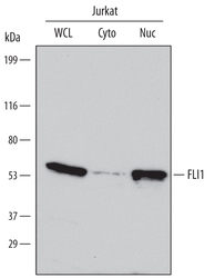
- Detection of Human FLI1 by Western Blot. Western blot shows lysates of Jurkat human acute T cell leukemia cell line. Gels were loaded with 30 µg of whole cell lysate (WCL), 20 µg of cytoplasmic (Cyto), and 10 µg of nuclear extracts (Nuc). PVDF Membrane was probed with 0.5 µg/mL of Human FLI1 Antigen Affinity-purified Polyclonal Antibody (Catalog # AF6474) followed by HRP-conjugated Anti-Sheep IgG Secondary Antibody (Catalog # HAF016). A specific band was detected for FLI1 at approximately 53 kDa (as indicated). This experiment was conducted under reducing conditions and using Immunoblot Buffer Group 1.
